Winter 2015 – Sticking to the Plan: Patient Preferences for Epidural Use During Labor

Sticking to the Plan: Patient Preferences for Epidural Use During Labor

Author: Lauren Ann Gamble1, Ashley Hesson1, Tiffany Burns2.

Author Affiliations:

1College of Human Medicine, Michigan State University, East Lansing, MI, USA

2Department of Family Medicine, Michigan State University, East Lansing, MI, USA

[button link=”http://msrj.chm.msu.edu/wp-content/uploads/2015/04/Winter-2015-Sticking-to-the-Plan.pdf” type=”big” color=”orange” newwindow=”yes”] Full Text Article PDF[/button]

Corresponding Author: Lauren Ann Gamble, gambleL2[at]msu.edu

Key Words: epidural; birth plan; labor analgesia; patient preference, decision making.

Abstract: Background: Women have been shown to value control in the labor experience, a desire that is often formalized into an explicit birth plan. Epidural preferences are a primary component of this plan. Despite this specification, women’s plans are not always carried out. This may be due to patient factors (e.g., dissatisfaction with labor), provider behaviors (e.g., frequent epidural offers), or situational variables (e.g., prolonged labor). Purpose: The current study investigates the relative impact of patient preference for epidural use as compared to provider suggestion and circumstances of labor. It hypothesizes that providing an epidural preference in a birth plan and receiving frequent epidural offers will predict epidural administration. Methods: Adult, postpartum women were surveyed about their labor experience at a high-volume obstetrics unit in a medium-sized community hospital. Responses to a structured survey instrument focused on prelabor preferences and labor characteristics. Descriptive statistics and multiple logistic regression modeling were used to analyze participant responses. Results: Eighty-three postlaboring women completed surveys, of which 79 surveys were analyzed. Eighty-four percent (N_66) received an epidural during their labor process, while 73% (N_58) desired an epidural as a part of their birth plan. Women were offered an epidural at a mean frequency of 0.2790.48 times per hour (median_0.14). The significant predictors of epidural administration were desire for an epidural in the birth plan (pB0.01) and the frequency of epidural offers (pB0.01). Wanting an epidural was associated with receiving an epidural. Conversely, increased frequency of being offered an epidural negatively correlated with epidural administration. Conclusions: Our findings indicate that personal preference is the most influential factor in determining whether or not a laboring woman will receive an epidural. Increasing provider attempts to offer an epidural – as represented by increased frequency of queries- decreased the likelihood that an epidural would be received.

Published on date: January 1, 2015

Senior Editor: Tina Chaalan

Junior Editor: Jennifer Monacelli

DOI: Pending

Citation: Gamble LA, Hesson A, Burns T. Sticking to the Plan: Patient Preferences for Epidural Use During Labor. Medical Student Research Journal. 2015;4(Winter):59-65.

References:


  1. Pilnick A, Dingwall R. On the remarkable persistence of asymmetry in doctor/patient interaction: a critical review. Soc Sci Med 2011; 72: 1374-82.
  2. Simkin P. Birth plans: after 25 years, women still want to be heard. Birth 2007; 34(1): 49-51.
  3. Namey EE, Lyerly AD. The meaning of ‘‘control’’ for childbearing women in the US. Soc Sci Med 2010; 71(4): 769-76.
  4. Miller AC, Shriver TE. Women’s childbirth preferences and practices in the United States. Soc Sci Med 2012; 75(4):709-16.
  5. Pennell A, Salo-Coombs V, Herring A, Spielman F, Fecho K. Anesthesia and analgesia_related preferences and outcomes of women who have birth plans. J Midwifery Women’s Health 2011; 56(4): 376-81.
  6. Horowitz ER, Yogev Y, Ben-Haroush A, Kaplan B. Women’s attitude toward analgesia during labor – a comparison between 1995 and 2001. Eur J Obstet Gynecol Reprod Biol 2004; 117(1): 30-32.
  7. Thompson R, Miller YD. Birth control: to what extent do women report being informed and involved in decisions about pregnancy and birth procedures? BMC Pregnancy Childbirth 2014; 14(1): 62.
  8. Toledo P, Sun J, Peralta F, Grobman WA, Wong CA, Hasnain-Wynia R. A qualitative analysis of parturients’ perspectives on neuraxial labor analgesia. Int J Obstet Anesth 2013; 22(2): 119-23.
  9. Fro¨ hlich S, Tan T, Walsh A, Carey M. Epidural analgesia for labour: maternal knowledge, preferences and informed consent. Irish Med J 2012; 104(10): 300-2.
  10. Pain relief during labor. ACOG committee opinion No 295. American College of Obstetricians and Gynecologists. Obstet Gynecol 2004; 104: 213.
  11. Johnson DE. Getting off the GoldVarb standard: introducing Rbrul for mixed-effects variable rule analysis. Lang Linguist Compass 2009; 3: 359-83.
  12. R Development Core Team. R: A language and environment for statistical computing [Computer program]. Vienna, Austria: R Foundation for Statistical Computing; 2012.
  13. Hadar E, Raban O, Gal B, Yogev Y, Melamed N. Obstetrical outcome in women with self-prepared birth plan. J Matern Fetal Neonatal Med 2012; 25(10): 2055-7.
  14. Hidaka R, Callister LC. Giving birth with epidural analgesia: the experience of first-time mothers. J Perinat Educ 2012; 21: 24.
  15. Kannan S, Jamison RN, Datta S. Maternal satisfaction and pain control in women electing natural childbirth. Reg Anesth Pain Med 2001; 26(5): 468-72.
  16. Hodnett ED. Pain and women’s satisfaction with the experience of childbirth: a systematic review. Am J Obstet Gynecol 2002; 186(5): S160-72.
  17. Lawrence HC III, Copel JA, O’Keeffe DF, Bradford WC, Scarrow PK, Kennedy HP, et al. Quality patient care in labor and delivery: a call to action. Am J Obstet Gynecol 2012; 207: 147-8.


[facebook] [retweet]

Winter 2015 – In Situ Thrombosis of the Pulmonary Arteries: An Emerging New Perspective on Pulmonary Embolism

In Situ Thrombosis of the Pulmonary Arteries: An Emerging New Perspective on Pulmonary Embolism

Author: Virginia Corbett1, Houria Hassouna2, Reda Girgis3

Author Affiliations:

1College of Human Medicine, Michigan State University, East Lansing, MI, USA

2Division of Thrombosis, Department of Internal Medicine, College of Human Medicine, Michigan State University, East Lansing, MI, USA

3Department of Pulmonary Medicine, College of Human Medicine, Michigan State University, Grand Rapids, MI, USA

[button link=”http://msrj.chm.msu.edu/wp-content/uploads/2015/04/Winter-2015-In-Situ-Thrombosis.pdf” type=”big” color=”green” newwindow=”yes”] Full Text Article PDF[/button]

Corresponding Author: Virginia Corbett, corbettv[at]msu.edu

Key Words: pulmonary embolism; in situ pulmonary artery thrombosis; deep vein thrombosis (DVT); pulmonary circulation; Virchow’s triad

Abstract: The annual incidence of pulmonary embolism(PE) in the United States is reported to be 0.69 per1,000 persons with mortality of up to 30% depending upon the size of the emboli.1 PE and deep venous thrombosis (DVT) are both considered manifestations ofthe same disease of venous thromboembolism. Virchowpostulated that dysfunction of vessel walls, alternationsin blood flow and hypercoagulability of theblood triggered inappropriate thrombus formation.2 DVT most commonly occurs as local clot formation in the deep calf veins. PE arises when clots break off from a peripheral DVT and become lodged within the pulmonary arterial vasculature. PE is routinely diagnosed when filling defects are found in the pulmonary arteries on computed tomography angiogram (CTA). Among the general population of patients presenting to emergency rooms, absence of DVT may occur in up to 57% of those diagnosed with PE.3 A high prevalence of isolated PE may suggest localized thrombus formation in the pulmonary arteries instead of embolization from peripheral clots.

Published on date: January 1, 2015

Senior Editor: Kailyne Van Stavern

Junior Editor: Garrett Roe

DOI: Pending

Citation: Corbett V. Hassouna H. Girgis R. In Situ Thrombosis of the Pulmonary Arteries: An Emerging New Perspective on Pulmonary Embolism . Medical Student Research Journal. 2015;4(Winter):54-8.

References:

  1. Office of the Surgeon General (US); National Heart, Lung, and Blood Institute (US). The surgeon general’s call to action to prevent deep vein thrombosis and pulmonary embolism. 2008. Available from: http://www.ncbi.nlm.nih.gov/books/NBK44181/ [cited 15 February 2014].
  2. Kyrle PA, Eichinger S. Deep vein thrombosis. Lancet 2005; 365(9465): 1163_74. doi: 10.1016/S0140-6736(05)71880-8
  3. Sohns C, Amarteifio E, Sossalla S, Heuser M, Obenauer S. 64-Multidetector-row spiral CT in pulmonary embolism with emphasis on incidental findings. Clin Imaging 2008; 32(5): 335_41. doi: 10.1016/j.clinimag.2008.01.028
  4. Van belle A, Bu¨ ller HR, Huisman MV, Huisman PM, Kaasjager K, Kamphuisen PW, et al. Effectiveness of managing suspected pulmonary embolism using an algorithm combining clinical probability, D-dimer testing, and computed tomography. JAMA 2006; 295(2): 172_9. doi: 10.1001/jama.295.2.172
  5. Kumar NG, Clark A, Roztocil E, Caliste X, Gillespie DL,Cullen JP. Fibrinolytic activity of endothelial cells from different venous beds. J Surg Res 2015; 194(1): 297_303. doi: 10.1016/j.jss.2014.09.028
  6. Rosenhek R, Korschineck I, Gharehbaghi-schnell E, Jakowitsch J, Bonderman D, Huber K, et al. Fibrinolytic balance of the arterial wall: pulmonary artery displays increased fibrinolytic potential compared with aorta. Lab Invest 2003; 83(6): 871_6. doi: 10.1097/01.LAB.0000073127.46392.9D
  7. Ryan US. Metabolic activity of pulmonary endothelium: modulations of structure and function. Annu Rev Physiol 1986; 48(1): 263_77. doi: 10.1146/annurev.ph.48.030186.001403.
  8. Goodman LR. Small pulmonary emboli: what do we know? Radiology 2005; 234(3): 654_8. doi: 10.1148/radiol.2343041326
  9. Ryan US. Structural bases for metabolic activity. Annu Rev Physiol 1982; 44(1): 223_39. doi: 10.1146/annurev.ph.44.030182.001255
  10. Key NS, Bach RR. Tissue factor as a therapeutic target. Thromb Haemost 2001; 85(3): 375_6. doi: 10.1517/14728222.6.2.159
  11. Martinelli I. Unusual forms of venous thrombosis and thrombophilia. Pathophysiol Haemost Thromb 2002; 32(5_6): 343_5. doi: 10.1159/000073595
  12. Agarwal PP, Wolfsohn AL, Matzinger FR, Seely JM, Peterson RA, Dennie C. In situ central pulmonary artery thrombosis in primary pulmonary hypertension. Acta Radiol 2005; 46(7): 696_700. doi: 10.1080/02841850500215501
  13. Russo A, De luca M, Vigna C, De Rito V, Pacilli M, Lombardo A, et al. Central pulmonary artery lesions in chronic obstructive pulmonary disease: a transesophagealechocardiography study. Circulation 1999; 100(17): 1808_15. doi: 10.1161/01.CIR.100.17.1808
  14. Wechsler RJ, Salazar AM, Gessner AJ, Spirn PW, Shah RM, Steiner RM. CT of in situ vascular stump thrombosis after pulmonary resection for cancer. AJR Am J Roentgenol 2001; 176(6): 1423_5. doi: 10.2214/ajr.176.6.1761423
  15. Lundy JB, Oh JS, Chung KK, Ritter JL, Gibb I, Nordmann GR, et al. Frequency and relevance of acute peritraumatic pulmonary thrombus diagnosed by computed tomographic imaging in combat casualties. J Trauma Acute Care Surg 2013; 75(2 Suppl 2): S215_20. doi: 10.1097/TA.0b013e318299da66
  16. Knudson MM, Gomez D, Haas B, Cohen MJ, Nathens AB. Three thousand seven hundred thirty-eight posttraumatic pulmonary emboli: a new look at an old disease. Ann Surg 2011; 254(4): 625_32. doi: 10.1097/SLA.0b013e3182300209
  17. Schulz C, Engelmann B, Massberg S. Crossroads of coagulation and innate immunity: the case of deep vein thrombosis. J Thromb Haemost 2013; 11 (Suppl 1): 233_41. doi: 10.1111/jth.12261
  18. Drake TA, Morrissey JH, Edgington TS. Selective cellular expression of tissue factor in human tissues. Implications for disorders of hemostasis and thrombosis. Am J Pathol 1989; 134(5): 1087_97.
  19. Polgar J, Matuskova J, Wagner DD. The P-selectin, tissue factor, coagulation triad. J Thromb Haemost 2005; 3(8): 1590_6. doi: 10.1111/j.1538-7836.2005.01373.x
  20. Sevestre MA, Quashie´ C, Genty C, Rolland C, Que´re´ I, Bosson JL, et al. Clinical presentation and mortality in pulmonary embolism: the Optimev study. J Mal Vasc 2010; 35(4): 242_9. doi: 10.1016/j.jmv.2010.05.004
  21. Van langevelde K, Flinterman LE, Van hylckama vlieg A, Rosendaal FR, Cannegieter SC. Broadening the factor V Leiden paradox: pulmonary embolism and deep-vein thrombosis as 2 sides of the spectrum. Blood 2012; 120(5):933_46. doi: 10.1182/blood-2012-02-407551
  22. De boer JD, Majoor CJ, Van’t veer C, Bel EH, Van der poll T. Asthma and coagulation. Blood 2012; 119(14): 3236_44. doi: 10.1182/blood-2011-11-391532
  23. Chung WS, Lin CL, Ho FM, Li RY, Sung FC, Kao CH, et al. Asthma increases pulmonary thromboembolism risk: a nationwide population cohort study. Eur Respir J 2014; 43(3):801_7. doi: 10.1183/09031936.00043313
  24. Majoor CJ, Kamphuisen PW, Zwinderman AH, Ten Brinke A, Amelink M, Rijssenbeek-Nouwens L, et al. Risk of deep vein thrombosis and pulmonary embolism in asthma. Eur Respir J 2013; 42(3): 655_61. doi: 10.1183/09031936.00150312
  25. Bertoletti L, Quenet S, Laporte S, Sahuquillo JC, Conget F, Pedrajas JM, et al. Pulmonary embolism and 3-month outcomes in 4036 patients with venous thromboembolism and chronic obstructive pulmonary disease: data from the RIETE registry. Respir Res 2013; 14: 75. doi: 10.1186/1465-9921-14-75
  26. Rizkallah J, Man SF, Sin DD. Prevalence of pulmonary embolism in acute exacerbations of COPD: a systematic review and metaanalysis. Chest 2009; 135(3): 786_93. doi:10.1378/chest.08-1516
  27. Konstantinides SV. Asthma and pulmonary embolism: bringing airways and vessels closer together. Eur Respir J 2014; 43(3): 694_6. doi: 10.1183/09031936.00009414
  28. Velmahos GC, Spaniolas K, Tabbara M, Abujudeh HH, de Moya M, Gervasini A, et al. Pulmonary embolism and deep venous thrombosis in trauma: are they related? Arch Surg 2009; 144(10): 928_32. doi:10.1001/archsurg.2009.97
  29. Van langevelde K, Sra´mek A, Vincken PW, Van rooden JK, Rosendaal FR, Cannegieter SC. Finding the origin of pulmonary emboli with a total-body magnetic resonance direct thrombus imaging technique. Haematologica 2013; 98(2):309_15. doi: 10.3324/haematol.2012.069195
  30. Kearon C, Akl EA. Duration of anticoagulant therapy for deep vein thrombosis and pulmonary embolism. Blood 2014; 123(12): 1794_1801. doi: 10.1182/blood-2013-12-512681
  31. PREPIC Study Group. Eight-year follow-up of patients with permanent vena cava filters in the prevention of pulmonary embolism: the PREPIC (Prevention du Risque d’Embolie Pulmonaire par Interruption Cave) randomized study. Circulation 2005; 112(3): 416_22. doi: 10.1161/CIRCULATIONAHA.104.512834
  32. Korkmaz A, Ozlu T, Ozsu S, Kazaz Z, Bulbul Y. Long-term outcomes in acute pulmonary thromboembolism: the incidence of chronic thromboembolic pulmonary hypertension and associated risk factors. Clin Appl Thromb Hemost 2012;18(3): 281_8. doi: 10.1177/1076029611431956

[facebook][retweet]

Winter 2015 – White Coat Sparty

White Coat Sparty.

Author:  Carter Anderson

Author Affiliations: College of Human Medicine, Michigan State University, East Lansing, MI, USA

[button link=”http://msrj.chm.msu.edu/wp-content/uploads/2015/04/Winter-2015-Cover-Art-Reflection.pdf” type=”big” color=”green” newwindow=”yes”] Full Text Article PDF[/button]

Corresponding author: Carter Anderson; carterbanderson[at]yahoo.com

Key Words: N/A

Abstract: Professional responsibility, compassion, honesty, respect for others, competence, and social responsibility are the characteristics that the Michigan State University College of Human Medicine strives to instill in every student.

Published on date: January 1, 2015

Senior Editor: N/A

Junior Editor: N/A

DOI: pending

Citation: Anderson C. White Coat Sparty. Medical Student Research Journal. 2015;4(Winter):52-53.

References: N/A


[facebook][retweet]

Winter 2015 – Letter from the Editors

Letter From the Editors.

Author: Jessica L Wummel1, Jack C Mettler2

Author Affiliations: 1College of Human Medicine, Michigan State University, East Lansing, MI, USA, 2College of Human Medicine, Michigan State University, Flint, MI, USA

[button link=”http://msrj.chm.msu.edu/wp-content/uploads/2015/04/Winter-2015-Letter-from-the-editors.pdf” type=”big” color=”green” newwindow=”yes”] Full Text Article PDF[/button]

Corresponding Author: Jessica L Wummel; Jessica[at]msrj.org, Jack C Mettler; Jack[at]msrj.org

Key Words: N/A

Abstract: The editors of MSRJ are excited to announce our Winter 2015 issue. As always, we were incredibly impressed by the caliber of submissions. This issue includes interesting articles written by medical students from UC Davis College of Medicine and Michigan State University College of Human Medicine.

Published on date: January 1, 2015

Senior Editor: N/A

Junior Editor: N/A

DOI: Pending

Citation: Wummel JL, Mettler JC. Letter From the Editors. Medical Student Research Journal. 2015;4(Winter):51.

References: N/A

 [facebook] [retweet]

Medical Students Create Art Depicting the Doctor-Patient Relationship

Each fall, the first year medical students at Michigan State University College of Human Medicine are asked to create an art project for their “Doctor/Patient Relationship” course. The students are assigned to reflect on what this relationship means and to both depict it and write a reflection about it. It’s from these pieces that we at MSRJ select our cover photos for each issue. There are so many amazing projects each year that we are unable to accommodate each one as a cover. We want to showcase these pieces to you. Below are some of these pieces.


P7
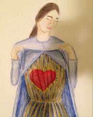
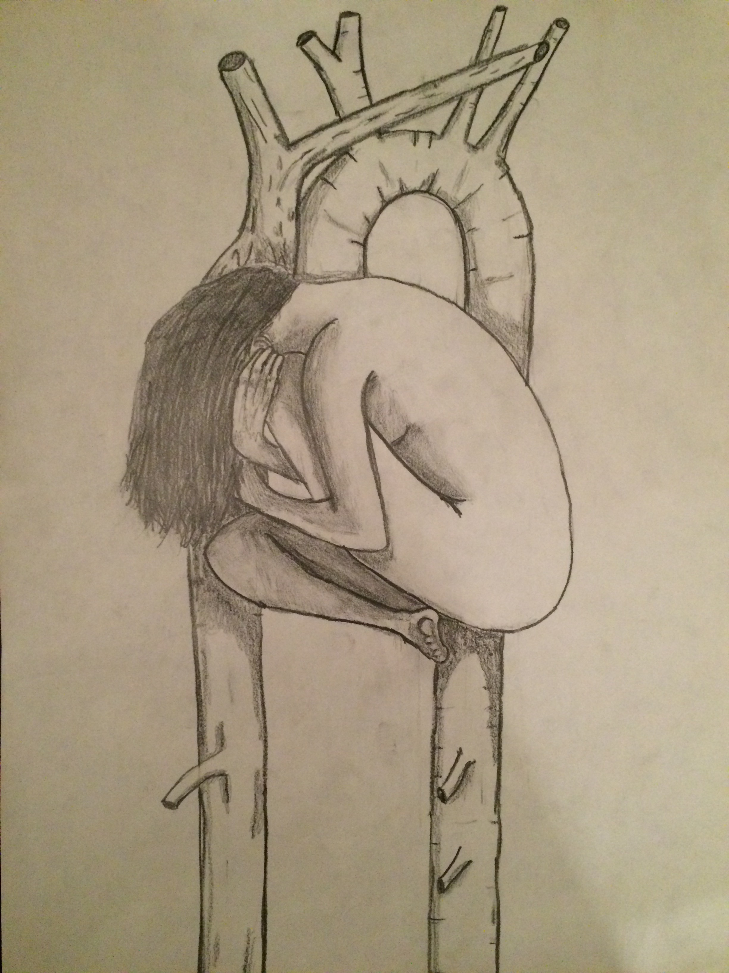
Created by: Jeremiah Reenders

When I started reflecting on the relationship between physicians and patients, I thought about the role of physicians and the function they serve for humanity. The long, arduous process of evolution has brought about infinitely complex, stunningly intricate, and beautifully balanced biological machinery that serves as the body in which we both perceive and interact with the universe around us. As impressive as our bodies are, however, there will always be enemies trying to hijack it, and innate imperfections that require the skill and wisdom of a healer to defend and restore our physical state. At first glance, it seems that this basic premise of our existence describes the role of physicians: to heal our bodies. Upon deeper reflection however, I couldn’t come to accept such a simplistic claim.

Somewhere in the course of our species’ existence, something was added that set us apart. The soul became entangled in every muscle fiber and between every neuron. Our existence became more than just the air entering and exiting our lungs and the blood that supplied our flesh. It transcended the physical and formed a connection between the human head, full of instinct and logic, and the heart, which breathes feeling in to an otherwise emotionless dimension. Although this connection is somewhat imperfect, it comprises the very core of who we are. It is impossible to tease apart the physical from the emotional, the mental from the chemical, the person and the flesh they live in. With my piece, I wanted to portray this fatal relationship. The heart of a person is a person. A physician should recognize that this person is far more complex than their anatomy and biochemistry. He/she should be prepared to look past his/her beliefs, values, and principles, and see only the person that exists before them. The patient is as much their soul as they are their body, and a physician must treat both.


IPPR_IWarner1
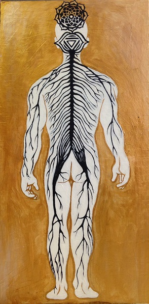
Created by: Irene Warner

For my Mid-Term Reflective Project, I decided to explore the concept of mindbody connection. While it may be tempting to reduce medicine to biochemical pathways and pure science, human beings are more complicated than the sum of their parts. When interacting with patients, it is important to remember that they are not only made up of quantifiable physical components, but of intangible spiritual and cultural components as well. My painting depicts a human nervous system fused with the Ajna (third-eye chakra) and the Sahasrara (crown chakra). The color gold in the background symbolizes perfection and balance, like the “golden mean” described by Aristotle in his philosophical teachings.

The nervous system is an essential part of human anatomy that coordinates all of the body’s function and sustains life. However, the concept of consciousness cannot be explained by nerves and synapses alone. In Hindu beliefs, the Ajna represents perception, intuition, and imagination. The Sahasrara is located at the crown of the head and represents higher consciousness, spirituality and wisdom. In my painting, the fusion of these symbols with the spinal cord and peripheral nervous system illustrates how the mind (i.e. higher consciousness and awareness) and physical body are extricably linked.

 “Heart of Patient Compassion”P1
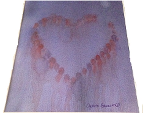
Created by: Jenna Bernson

I want to bring compassion to the patient-physician relationship. Compassion is represented by the heart shape as well as by the use of the color red. The heart, symbolic of my heart, is formed by abstract figures which stand for my patients. Notice that the heart is not complete, there is a portion left open. This represents always leaving an open heart to care for more patients. The background is done in purple, a cool color. This represents a sense of calmness I want to have and to give to my patients despite the fact that they may be going through very turbulent times or even though outside of our relationship, the world may be bleak (as represented by the starkness of the purple background). The figures are shown in both red and purple and every color in between, representing an openness of the heart also for diversity and inclusion. Culture influences so much of a person, from their daily activities to their approach to healthcare. So, understanding a patient’s values and culture is integral for a good patient-physician relationship. The differing perspectives and viewpoints of the patients is symbolized by the fact that every figure is given a different place in the heart. Each figure would see something slightly different even though they are all part of the same picture. Likewise, my own culture and values will influence the way in which I live and interact, even in my future practice. Therefore, I must be aware of my own background, beliefs and perspective to try to minimize and be aware of possible implicit biases. My perspective is represented by the use of abstract technique. Every person looking at this piece will pick out slightly different things that they see or will take away, which is undoubtedly different from what I see.

The Hospital Gown: Exploring the Patient-Physician Relationship Through Art and Metaphor

P2Created by: Carina Mendoza

My project illustrates (through art) a typical patient, with a hospital gown covering the body like an inanimate protective barrier: shielding the host from an oblivious and, perhaps, indifferent external world. The hospital gown is symbolic of an anonymous person (in this case a female) who enters a physician’s office; the office of a healer who is also dressed in a gown (but a gown of identity, authority and distinction), and who may be unaware of the inherent role that the respective gowns play in the patient-physician relationship, or of the concerned heart that beats beneath the personal garments, veil of skin and cage of bones (the body) of the anonymous female. The hospital gown reminds me of several instructional and significant tenets of our profession as healers. First, I am reminded that it is our responsibility to remain patient-centered, to facilitate the patient-physician relationship, to remove any real or imagined inhibiting barriers created by the hospital gowns, and to establish a trustworthy, caring environment where the seemingly anonymous gown draped-person becomes a client, a patient with a name and feelings, and an individual with desires to be cared for as a deserving, respected and dignified human being. Second, I am reminded that as physicians we must resist any inclination to become oblivious, dismissive or disinterested in the role that gender, culture, religion and other non-physical factors play in the healing processes: we must, instead, remain cognizant of the connection between mind and body, of facial gestures and body language (ours and the patient’s); and about our own gender, cultural and religious biases that may obstruct our ability to remain empathic and sensitive to the plethora of insecurities that accompany pain and suffering. Third, I am reminded of the kindred human connection that we have with our patients, and that at some point in our lives we may experience a role reversal and become victims of disease and illness; yearning for the same sensitivity, compassion, consideration and understanding that we may be (unwittingly) denying our patients.

“RED Bond”

P12
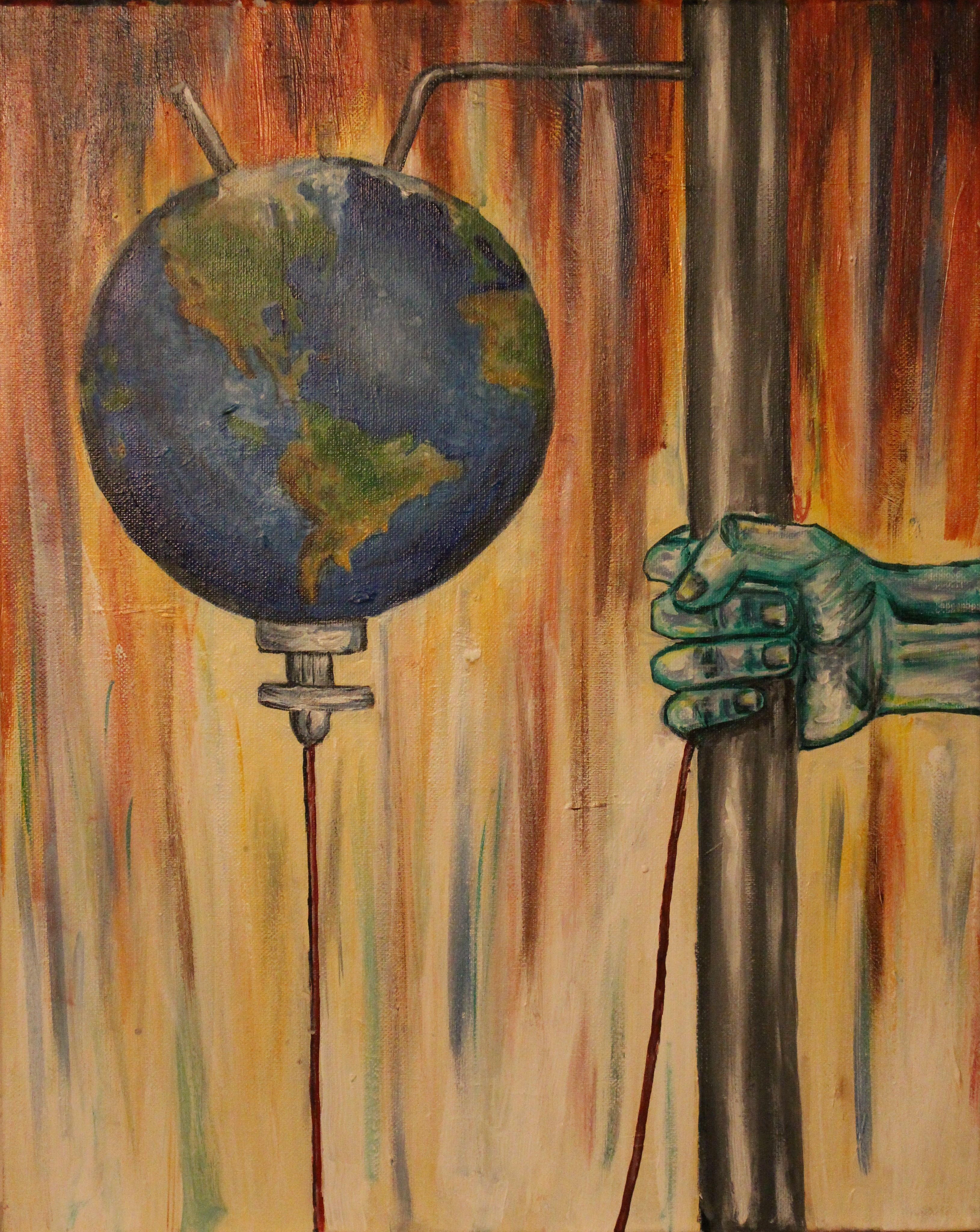
Created by: Abdelouahid Souala

How can health care practitioners provide culturally competent care? Patients can present from a wide variety of backgrounds and cultures. Must we be aware of each culture’s customs in order to provide culturally competent care? I initially thought this is how cultural competence is practiced. However, I quickly realized that this is not feasible. Not only is there a great variety of cultures, but cultures evolve with time and new cultures and customs are formed frequently. The solution is quite simple, focus on the patient.

Each patient is unique. Culture is only one aspect of the patient. Nonetheless, it can be a very important aspect in some patients that helps guide their everyday life. Cultural competence is achieved by connecting with patients and asking about their cultural needs. Furthermore, collaboration with patient on how to meet these needs will help the provider incorporate specific interventions tailored to meet the patient’s cultural needs. Sometimes, these needs are easily met and are incorporated smoothly into the plan of care. Other times, these needs can be very challenging to meet. This is especially true when the cultural needs pose an obstacle to the planned care or are completely opposed to the standard medical treatment. This is one it’s most important to remember to keep the focus on the patient. If these challenging cultural needs are neglected with the intention of providing the scientifically deemed best therapy for the disease, then only the disease is being cared for and the rest of the patient is neglected. Sometimes this means that health care professionals must neglect the disease in order to care for the patient.

The painting is a great reminder of how to deliver culturally competent care. I made it to help remind me of cultural competence when I start practicing medicine. Healthcare professionals must act like the IV in the painting. Connect with the patient, identify their cultural needs, prepare a medical therapy that fulfills these needs, and finally deliver this personalized therapy to the patient.


P9
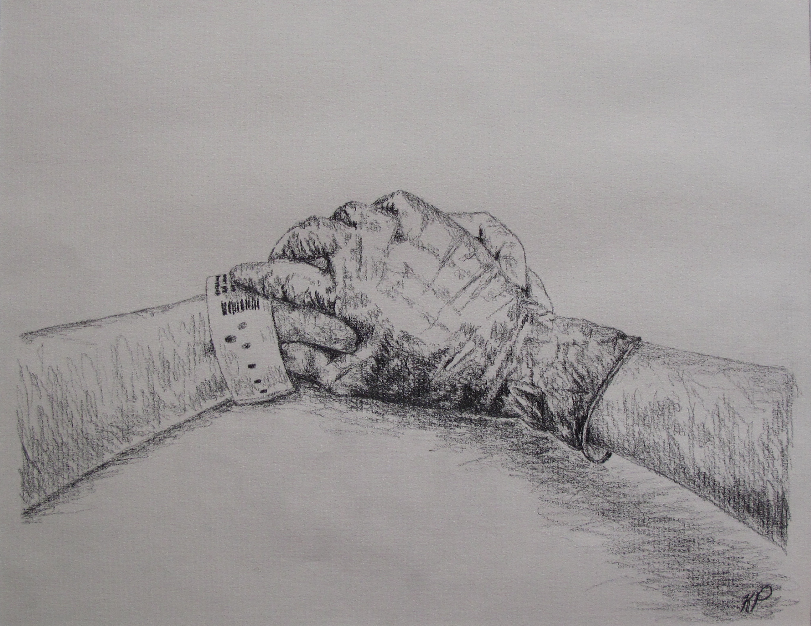
Created by: Kristina Priessnitz

I can remember the intensity of her face as the operating room nurses wheeled the patient’s bed through the O.R. doors, her wide eyes haunting me even a year from now. I was a research assistant for Obstetric Anesthesiology, and I was upstairs in Labor and Delivery with my coworker to measure blood loss in C-section cases with our newly created calculation worksheet. Before we entered the room to follow the patient, our senior research supervisor and attending anesthesiologist took my coworker and me aside to talk in the hallway.

“You will not be measuring blood loss in this case, but just observing. This woman is here for a D&C; her baby had lethal genetic mutations and malformations,” she said. “Understandably she is quite upset. She wanted to have this baby.”

My mind was spinning. A D&C procedure, a dilation and curettage, was generally used in induced, therapeutic, or incomplete abortions and miscarriages. I could not imagine the turmoil and emotion that must have been coursing through the patient; I already caught a glimpse as she passed by earlier.

Walking into the O.R. behind the attending anesthesiologist, my coworker and I watched the nurses and doctors work in solemn silence. They carefully lifted her onto the operating table and gently laid her arms on the outstretched, horizontal armrests. As the anesthesiology residents worked to clip the blue drape curtain, and hook up patient monitors and IVs, I couldn’t help but watch the patient. Her eyes focused to the ceiling as she bit her lower lip, trying to control her expression. However, despite her effort, small clear tears slowly started to stream down her face sideways. So desperately, I wanted to reach out to her and comfort her, but my coworker and I were supposed to be flies on the wall—no touching the patient. Then the attending anesthesiologist did something I will never forget: she walked around the table to face the patient, ignoring the residents working above the patient’s head. The attending clasped the patient’s hand with her gloved hand and murmured something soothing to the patient while she wiped the patient’s tears. Once the patient was calmer, she unfolded a warm blanket over patient’s chest and outstretched arms while rubbing the patient’s arms like a loving mother would to a sleepless child. The patient slowly closed her eyes and drifted asleep.

Reflecting back as a medical student now, the powerful imagery I witnessed that day is still seared to my brain: a physician’s gloved hand clasping the outstretched patient’s hand. It was such a simple, but meaningful gesture of the physician’s compassion toward the frightened patient. As physicians, we are privileged to serve patients when they need us most. Often times, that may mean encountering patients on some of their most difficult, challenging, or life-changing moments. Although the attending anesthesiologist has undoubtedly encountered many D&C procedures in her lifetime of work, I admired how in tune she remained to the patient and her needs. She recognized how difficult this operation was for the patient and provided a hand to hold and soothing words when the patient was most vulnerable.

Within the career of medicine, we are long-standing witnesses to the great highs and lows of life. Physicians are not only healers of the body, but of the mind and soul as well. Never must this work become so routine that we lose sight of humanity in our daily efforts. Never must we forget to hold our fellow brother or sister’s hand.


P11
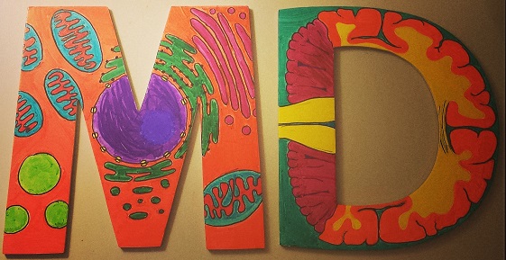
Created by: Jessica Priestley

The theme of my creative project for this course was “the art and science of medicine” – a concept that has made an enormous impact on both how I envision my career as a physician, and the relationships I will have with my patients.

The base of my project are two cardboard letters – M and D. Those letter reflect both the culmination of a dream to come to (and complete) medical school, and the commencement of a career. More than that, I believe that the M.D. degree represents a part of both who I am and who I want to be. The degree represents a tremendous amount of hard work for me, personally. However, and more importantly, it signifies the special trust placed in medical professionals by the public. It signifies the type of person who dedicates her life to the service of others.

I’ve drawn certain steps in the organizational hierarchy of life on each face of the two letters. Starting on one face of the “M,” there is a single cell with its various organelles: a nucleus in the center with its nucleolus and nuclear pores, surrounded to one side by rough endoplasmic reticulum. The functions of that cell are integral to the functioning of the tissue, organ, and organism as a whole. As a result, a great deal of medical attention is paid to the cellular level of a patient – from specific receptor blockers to chemotherapeutics that inhibit transcription, we rely on our ability to manipulate cellular function in order to treat disease in a whole patient. However, it’s not enough for physicians to spend their time thinking about cells and organelles.

On the first face of the “D” is a histological depiction of nervous tissue, featuring axons branching away from a neuronal cell body and nearby glial cells. Physicians must expand their consideration of disease and health to consider the function of tissues that cells comprise. I picked nervous tissue because I think it nicely illustrates the fact that one cannot focus too heavily on a single cell type (neurons, for example) without considering the role of “supporting” cells (glia, for example). Historically, the scientific world has put a lot of energy into studying neurons because they seem synonymous with nervous tissue. Today, we are learning more and more about the critical role glial cells play in the function and dysfunction of the nervous system.

Turning over the “D,” I’ve drawn a coronal section of a brain, the whole organ comprised by nervous tissue. To this point, my illustrations have dealt with the basic science of medicine – the cellular biology, histology, anatomy, and physiology. That information is the foundation of our eventual medical practice – this is the science of medicine.

The art of medicine is less tangible, and is represented both by my depiction of the cell, tissue, and brain as caricatures of the “real thing,” as well as by my abstraction on the flip side of the “M.” Accompanying the illustration are the three virtues (courage, humility and mercy) and six professional responsibilities (compassion, competence, honesty, professional responsibility, respect, and social responsibility) from the Michigan State University College of Human Medicine “Virtuous Professional” document. Each of those qualities contribute to the skill of practicing the “art of medicine,” which to me means the ability for a physician to relate and respond to a patient’s needs as both a collection of cells, tissues, and organs, and as a living, feeling human being with medical needs that will surely traverse the divide between biology, psychology, and sociology. While cells, tissues, and organ systems represent “doctor-centered-ness,” these qualities are what comprise “patient-centered-ness” and solidify the physician-patient relationship as more meaningful and significant than a simple business transaction. We spent some time in class talking about those values in a very black-and-white sense, as if they are easily attained and acquired after an hour’s discussion. Instead, those words rest on a colorful background representing the richness of experiences that both we as physicians and our patients bring to the medical experience. These concepts are not clear cut, and will not always be easy. There will be mistakes, but there will (hopefully) also be growth from those mistakes.

Finally, my project has the physical quality of dimension (i.e. both sides of the letters), meant to underscore the complexities of the physician-patient relationship. Those complexities are multi-dimensional and include some themes from this course: a diverse patient population with unique cultural, ethnic, and religious beliefs about both wellness and illness, patient autonomy, fiduciary relationships and the dynamics of trust and power, and the flow of information as doctors inform their patients of options, risks, and rewards. It is that richness that I look forward to most as I advance through my training because it is what makes the illness of Mrs. Abernathy with heart failure a different challenge than Mr. Brown with heart failure, even though the molecular/cellular aspects of their shared disease might be quite similar.


P6
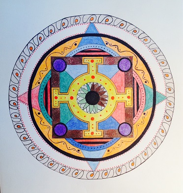
Created by: Dan Roberts

As I descended into impassable rivers I no longer felt guided by the ferrymen…

_arthur rimbaud

A mandala (Sanskrit: ‘circle’) is a design, typically seen in Hindu and Buddhist contexts, symbolizing the universe. Tibetan Buddhist monks often use mandalas as a meditation tool, and Native American Indians and various groups use mandalas in rituals for healing. The Swiss psychoanalyst Carl Jung utilized mandalas with his patients, as he viewed the mandala as a tool to explore the human psyche.

So, as this project was intended to be reflective, my immediate thought was that I would utilize the mandala as part of my own reflective process. The center of my mandala represents my eye (my right eye has partial heterochromia) looking back at me—to remind me of my own vulnerabilities, as well as the importance of being aware of my own values, biases, etc., and the effect these programs can have on my future relationships with patients.

Prior to coming to medical school, I worked with clients struggling with mental distress, as well as addiction issues. I very much enjoyed working in this capacity, but felt that I needed to continue on with medical training, to allow me to better promote healing through a mind-body connection. As this moment, I foresee myself pursuing psychiatry, so that I can help individuals to navigate those impassible rivers to find their own inner healing (represented in my mandala by the dark black ring encompassing an inner array of color and light).

The doctor is effective only when he himself is affected. Only the wounded physician heals.

_carl jung


P5
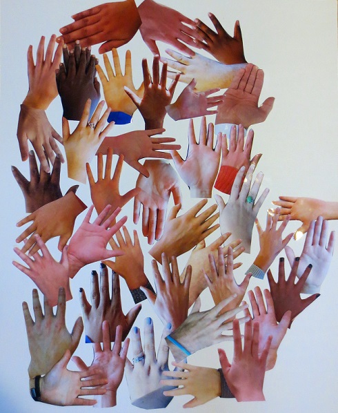
Created by: Felicia Nip

Each hand is a reminder that we, as future physicians, hold a power to lead and guide the health of our patients. But, hands are meant to give as much as they are meant to receive. Therefore, we must humbly allow our patients to guide us in using our powerful tools. Each hand is also a reminder to acknowledge diversity. We may understand our own capabilities, culture, experiences, and ideas, but we must also respect and attempt to understand both our colleagues’ and patients’ as well.

On display are hands from MSU CHM (M1) students. Every one of us will bring diverse cultural, educational, religious, and geographical experiences across Michigan communities in our third and fourth years. The challenge I pose for my peers, and myself is to think about what can, should, and will be accomplished with our hands each year. I believe each pair of hands can and will change innumerable lives.  My goal as a future physician is to wear out my hands by serving others.

[facebook]

[retweet]

MSRJ Elective Ping-Pong Tournament

Each spring the MSRJ organizes an elective for students interested in learning how to critically evaluate research. As a part of the elective we invite guest speakers to speak to us about the importance of research and have interactive sessions on how to act as peer reviewers for articles submitted to our journal. To raise awareness of the upcoming elective and promote interest in our journal, the MSRJ hosted a ping-pong tournament/pizza party in both the East Lansing and Grand Rapids Campus.  The winner in Grand Rapids was Cory Messingschlager and the winner in East Lansing was former MSU tennis star Christian Roehmer. Both guys won a $25 gift card to Starbucks, an official ping-pong trophy, and most importantly bragging rights.

East Lansing

  20141201_172847

20141201_182949

20141201_190846

Grand Rapids

 GR2

[retweet] [facebook]

Fall 2014 – The Growth of Medical Student Opportunities in Global Health

The Growth of Medical Student Opportunities in Global Health.

Author: Johnathan Kao, MPH

Author Affiliations: College of Human Medicine, Michigan State University, Flint, MI, USA

[button link=”http://msrj.chm.msu.edu/wp-content/uploads/2014/12/Fall-2014-Editorial-Global-Health.pdf” type=”icon” icon=”download” color=green] Full Text Article PDF[/button]

Corresponding Author: Johnathan Kao; johnathan.kao[at]msrj.org

Key Words: sexual health; relationships; intimacy; radiotherapy; psycho-supportive treatment; hormone therapy.

AbstractSince the establishment of the World Health Organization on April 7, 1948,1 global health has grown in prominence and popularity among health care workers at all levels of training. International clinical rotation electives have been available to students for over half a century2 and interest in these programs has risen steadily over the decades. During this period, many organizations established programs for students and faculty interested in global health research and service. In 2006, these organizations united under the WHO’s Global Health Workforce Alliance to assist students and faculty in becoming more involved in global health activities.3 Despite these Changes, in 2007, Drain et al recognized a lack of global health education in medical schools and growing student interest, calling for more opportunities to fill the gap.4

Published on date: September 31, 2014

Senior Editor: N/A

Junior Editor: N/A

DOI: Pending

Citation: Kao J. The Growth of Medical Student Opportunities in Global Health. Medical Student Research Journal. 2014;4(Fall):48-50.

References:

  1. World Health Organization. History of WHO. 2014. Accessed from: http://www.who.int/about/history/en/ [cited 21 June 2014].
  2. Bissonette R, Route C. The Educational Effect of Clinical Rotations in Nonindustrialized Countries. Fam Med 1994; 26(4):226-31.
  3. World Health Organization. Global Health Education Consortium. 2014. Accessed from: http://www.who.int/workforcealliance/members_partners/member_list /ghec/en/ [cited 21 June 2014].
  4. Drain PK, Primack A, Hunt DD, Fawzi WW, Holmes KK, Gardner P. Global Health in Medical Education: A Call for More Training and Opportunities. Acad Med 2007; 82(3):226-30. doi: 10.1097/ACM.0b013e3180305cf9
  5. Hag C, Rothenberg D, Gjerde C, Bobula J, Wilson C, Bickley L, Cardelle A, Joseph A. New World Views: Preparing Physicians in Training for Global Health Work. Fam Med 2000; 32(8):566-72.
  6. Medical School Graduation Questionnaire: All School Summary Report. 2013. Accessed from: https://www.aamc.org/download/350998/data/2013 gqallschoolssummaryreport.pdf [cited 21 June 2014].
  7. Imperato PJ. A Third World International Health Elective for U.S. Medical Students: The 25-year Experience of the State University of New York, Downstate Medical Center. J Community Health 2004; 29(5):337-73. doi:10.1023/b:johe.0000038652.65641.0d
  8. Pust RE, Moher SP. A Core Curriculum for International Health: Evaluating Ten Years’ Experience at the University of Arizona. Acad Med 1992; 67(2):90-4. doi:10.1097/00001888-199202000-00007
  9. Haq C, Rothenberg D, Gjerde C, Bobula J, Wilson C, Bickley L, Cardelle A, Joseph A. New World Views: Preparing Physicians in Training for Global Health Work. Fam Med 2000; 32(8):566-72.
  10. Suchdev P, Ahrens K, Click E, Macklin L, Evangelista D, Graham E. A Model for Sustainable Short-Term International Medical Trips. Ambul Pediatr 2007; 7(4):317-20. doi: 10.1016/j.ambp.2007.04.003
  11. Montgomery LM. Short-Term Medical Missions: Enhancing or Eroding Health? Missiology 1993; 21(3):333-41. doi: 10.1177/009182969302100305
  12. Crump JA, Sugarman J, Working Group on Ethics Guidelines for Global Health Training (WEIGHT). Ethics and Best Practice Guidelines for Training Experiences in Global Health. Am J Trop Med Hyg 2010; 83(6):1178-82. doi: 10.4269/ajtmh.2010.10-0527
  13. AAMC GSA Steering Committee. Guidelines for Premedical and Medical Students Providing Patient Care During Clinical Experiences. AAMC 2011. Accessed from: https://www.aamc.org/download/181690/data/guidelinesforstudentsprovidingpatientcare.pdf [cited 21 June 2014].
  14. DeCamp M, Enumah S, O’Neill D, Sugarman J. Perceptions of a Short-Term Medical Programme in the Dominican Republic: Voices of Care Recipients. Glob Public Health 2014; 9(4):411-25. doi: 10.1080/17441692.2014.893368
  15. Accreditation Council of Graduate Medical Education. Common Program Requirements. ACGME 2013. Accessed from: https://www.acgme.org/ acgmeweb/Portals/0/PFAssets/ProgramRequirements/CPRs2013.pdf [cited 21 June 2014].
  16. Thompson MJ, Huntington MK, Hunt DD, Pinsky LE, Brodie JJ. Educational Effects of International Health Electives on U.S. and Canadian Medical Students and Residents: A Literature Review. Acad Med 2007; 82(3):226-30. doi: 10.1097/00001888-200303000-00023

[facebook] [retweet]

Fall 2014 – Comparing Current Screening Modalities for Colorectal Cancer and Precancerous Lesions: Is Colonoscopy the Method of Choice?

Comparing Current Screening Modalities for Colorectal Cancer and Precancerous Lesions: Is Colonoscopy the Method of Choice?

Author: Puneet K. Singh

Author Affiliations: Saba University School of Medicine, Saba, Dutch Caribbean

[button link=”http://msrj.chm.msu.edu/wp-content/uploads/2014/12/Fall-2014-CRC-Screening.pdf” type=”icon” icon=”download” color=green] Full Text Article PDF[/button]

Corresponding Author: Puneet K. Singh; pun33t.singh[at]gmail.com

Key Words: Colonoscopy; colorectal neoplasms; sigmoidoscopy; CT colonography; mass screening.

Abstract: Colorectal cancer (CRC) is the third most common form of cancer and the second leading cause of cancer death in the Western world. Presently, screening tools such as colonoscopy, sigmoidoscopy, fecal occult blood test (FOBT) and computed tomographic colonography (CTC) are available for CRC screening. The debate over which screening tool is most effective in detecting CRC and precancerous lesions is ongoing. Many recent studies have identified colonoscopy as the most sensitive and specific screening modality for CRC. However, a number of factors have prevented colonoscopy from being widely accepted. Less invasive techniques such as sigmoidoscopy and CTC are growing in popularity among physicians and patients who are apprehensive about colonoscopy screening; although many still are yet to experience the procedure first-hand. This literature review will attempt to validate the growing theory that colonoscopy is superior to other modalities for the diagnosis and screening of CRC and reduces the risk of CRC mortality. In order to do so, the paper will compare the risks and benefits of colonoscopy to sigmoidoscopy and CTC. It will further look at the different aspects that encompass a patient’s decision to partake in screening, such as basic knowledge about CRC, history of CRC in the family, advice from physicians and individual beliefs about what screening entails. Finally, this paper will propose ways in which colonoscopy screening can be improved and thus surpass other screening modalities to universally become the first choice for CRC screening.

Published on date: September 31, 2014

Senior Editor: Jack Mettler

Junior Editor: David Carr

DOI: Pending

Citation: Singh PK. Comparing Current Screening Modalities for Colorectal Cancer and Precancerous Lesions: Is Colonoscopy the Method of Choice? Medical Student Research Journal. 2014;4(Fall):34-47.

References:

  1. Holt PR, Kozuch P, Mewar S. Colon cancer and the elderly: from screening to treatment in management of GI disease in the elderly. Best Pract Res Clin Gastroenterol. 2009;23:889-907. doi: 10.1016/j.bpg.2009.10.010
  2. Siegel R, Naishadham D, Jemal A. Cancer statistics, 2012. CA Cancer J Clin. 2012;62:10-29. doi:  10.3322/caac.20138
  3. Manne U, Shanmugam C, Katkoori VR, Bumpers HL, Grizzle WE. Development and progression of colorectal neoplasia. Cancer Biomark. 2010;9:235-265. doi: 10.3233/CBM-2011-0160
  4. Qaseem A, Denberg TD, Hopkins RH, Jr., et al. Screening for colorectal cancer: a guidance statement from the American College of Physicians. Ann Intern Med. 2012;156:378-386. doi: 10.7326/0003-4819-156-5-201203060-00010
  5. Centers for Disease C, Prevention. Vital signs: Colorectal cancer screening, incidence, and mortality–United States, 2002-2010. MMWR Morb Mortal Wkly Rep. 2011;60:884-889.
  6. Lieberman D. Colorectal cancer screening: practice guidelines. Dig Dis. 2012;30 Suppl 2:34-38. doi: 10.1159/000341891
  7. Centers for Disease C, Prevention. Vital signs: colorectal cancer screening test use – United States, 2012. MMWR Morb Mortal Wkly Rep. 2013;62:881-888.
  8. Kahi CJ, Anderson JC, Rex DK. Screening and surveillance for colorectal cancer: state of the art. Gastrointest Endosc. 2013;77:335-350. doi: 10.1016/j.gie.2013.01.002
  9. Rabeneck L, Paszat LF, Saskin R, Stukel TA. Association between colonoscopy rates and colorectal cancer mortality. Am J Gastroenterol. 2010;105:1627-1632. doi: 10.1038/ajg.2010.83
  10. Manser CN, Bachmann LM, Brunner J, Hunold F, Bauerfeind P, Marbet UA. Colonoscopy screening markedly reduces the occurrence of colon carcinomas and carcinoma-related death: a closed cohort study. Gastrointest Endosc. 2012;76:110-117. doi: 10.1016/j.gie.2012.02.040
  11. Singh H, Nugent Z, Demers AA, Kliewer EV, Mahmud SM, Bernstein CN. The reduction in colorectal cancer mortality after colonoscopy varies by site of the cancer. Gastroenterology. 2010;139:1128-1137. doi: 10.1053/j.gastro.2010.06.052
  12. Baxter NN, Goldwasser MA, Paszat LF, Saskin R, Urbach DR, Rabeneck L. Association of colonoscopy and death from colorectal cancer. Ann Intern Med. 2009;150:1-8.
  13. Bretagne JF, Hamonic S, Piette C, et al. Variations between endoscopists in rates of detection of colorectal neoplasia and their impact on a regional screening program based on colonoscopy after fecal occult blood testing. Gastrointest Endosc. 2010;71:335-341. doi: 10.1016/j.gie.2009.08.032
  14. Adler A, Wegscheider K, Lieberman D, et al. Factors determining the quality of screening colonoscopy: a prospective study on adenoma detection rates, from 12,134 examinations (Berlin colonoscopy project 3, BECOP-3). Gut. 2013;62:236-241. doi: 10.1136/gutjnl-2011-300167
  15. Baxter NN, Warren JL, Barrett MJ, Stukel TA, Doria-Rose VP. Association between colonoscopy and colorectal cancer mortality in a US cohort according to site of cancer and colonoscopist specialty. J Clin Oncol. 2012;30:2664-2669. doi: 10.1200/JCO.2011.40.4772
  16. Ko CW, Dominitz JA, Green P, Kreuter W, Baldwin LM. Specialty differences in polyp detection, removal, and biopsy during colonoscopy. Am J Med. 2010;123:528-535. doi: 10.1016/j.amjmed.2010.01.016
  17. Graser A, Stieber P, Nagel D, et al. Comparison of CT colonography, colonoscopy, sigmoidoscopy and faecal occult blood tests for the detection of advanced adenoma in an average risk population. Gut. 2009;58:241-248. doi: 10.1136/gut.2008.156448
  18. Schoen RE, Pinsky PF, Weissfeld JL, et al. Colorectal-cancer incidence and mortality with screening flexible sigmoidoscopy. N Engl J Med. 2012;366:2345-2357. doi: 10.1056/NEJMoa1114635
  19. Hoff G, Grotmol T, Skovlund E, Bretthauer M, Norwegian Colorectal Cancer Prevention Study G. Risk of colorectal cancer seven years after flexible sigmoidoscopy screening: randomised controlled trial. BMJ. 2009;338:b1846. doi: 10.1136/bmj.b1846
  20. Johnson CD, Chen MH, Toledano AY, et al. Accuracy of CT colonography for detection of large adenomas and cancers. N Engl J Med. 18 2008;359:1207-1217. doi: 10.1056/NEJMoa0800996
  21. Zalis ME, Blake MA, Cai W, et al. Diagnostic accuracy of laxative-free computed tomographic colonography for detection of adenomatous polyps in asymptomatic adults: a prospective evaluation. Ann Intern Med. 2012;156:692-702. doi: 10.7326/0003-4819-156-10-201205150-00005
  22. Atkin W, Dadswell E, Wooldrage K, et al. Computed tomographic colonography versus colonoscopy for investigation of patients with symptoms suggestive of colorectal cancer (SIGGAR): a multicentre randomised trial. Lancet. 2013;381:1194-1202. doi: 10.1016/S0140-6736(12)62186-2
  23. von Wagner C, Ghanouni A, Halligan S, et al. Patient acceptability and psychologic consequences of CT colonography compared with those of colonoscopy: results from a multicenter randomized controlled trial of symptomatic patients. Radiology. 2012;263:723-731. doi: 10.1148/radiol.12111523
  24. de Wijkerslooth TR, de Haan MC, Stoop EM, et al. Reasons for participation and nonparticipation in colorectal cancer screening: a randomized trial of colonoscopy and CT colonography. Am J Gastroenterol. 2012;107:1777-1783. doi:  10.1038/ajg.2012
  25. de Wijkerslooth TR, de Haan MC, Stoop EM, et al. Burden of colonoscopy compared to non-cathartic CT-colonography in a colorectal cancer screening programme: randomised controlled trial. Gut.2012;61:1552-1559. doi:  10.1038/ajg.2012
  26. Courtney RJ, Paul CL, Sanson-Fisher RW, et al. Individual- and provider-level factors associated with colorectal cancer screening in accordance with guideline recommendation: a community-level perspective across varying levels of risk. BMC Public Health. 2013;13:248. doi: 10.1186/1471-2458-13-248
  27. Fenton JJ, Jerant AF, von Friederichs-Fitzwater MM, Tancredi DJ, Franks P. Physician counseling for colorectal cancer screening: impact on patient attitudes, beliefs, and behavior. J Am Board Fam Med. 2011;24:673-681. doi: 10.3122/jabfm.2011.06.110001

Fall 2014 – Alzheimer’s Disease: A Clinical and Basic Science Review

Alzheimer’s Disease: A Clinical and Basic Science Review. 

Author: Igor O. Korolev

Author Affiliations: College of Osteopathic Medicine and Neuroscience Program, Michigan State University, East Lansing, MI, USA

[button link=”http://msrj.chm.msu.edu/wp-content/uploads/2014/12/Fall-2014-Alzheimers-Disease.pdf” type=”icon” icon=”download” color=green] Full Text Article PDF[/button]

Corresponding Author: Igor O. Korolev; korolevi[at]msu.edu

Key Words: Alzheimer’s disease; mild cognitive impairment; dementia; neurodegeneration; neuroimaging; biomarkers.

Abstract: Alzheimer’s disease (AD) is the most common cause of dementia in older adults and an important public health problem. The purpose of this review article is to provide a brief introduction to AD and the related concept of mild cognitive impairment (MCI). The article emphasizes clinical and neurobiological aspects of AD and MCI that medical students should be familiar with. In addition, the article describes advances in the use of biomarkers for diagnosis of AD and highlights ongoing efforts to develop novel therapies.

Published on date: September 31, 2014

Senior Editor: Liza Gill

Junior Editor: Timothy Smith

DOI: Pending

Citation: Korolev IO. Alzheimer’s Disease: A Clinical and Basic Science Review.  Medical Student Research Journal. 2014;4(Fall):24-33.

References:

  1. Alzheimer A. About a Peculiar Disease of the Cerebral Cortex. Allg Z Psychiatr. 1907; 64: 146-148.
  2. Alzheimer A. About a Peculiar Disease of the Cerebral Cortex. Alzheimer Dis Assoc Disord. 1987; 1: 3-8.
  3. Maurer K, Volk S, Gerbaldo H. Auguste D and Alzheimer’s Disease. Lancet. May 1997; 349(9064): 1546-1549. doi: 10.1016/S0140-6736(96)10203-8
  4. Gilman, S. Oxford American Handbook of Neurology. Oxford, England: Oxford University Press; 2010.
  5. McKhann GM, Knopman DS, Chertkow H, Hyman BT, Jack CR, et al. The diagnosis of dementia due to Alzheimer’s disease: Rocommendations from the National Institute on Aging-Alzheimer’s Association workgroups on diagnostic guidelines for Alzheimer’s disease. Alzheimers Dement. May 2011; 7(3): 263-269. doi: 10.1016/j.jalz.2011.03.005
  6. Shadlen M, Larson E (2012) Evaluation of cognitive impairment and dementia. In: Basow D, editor. UpToDate. Waltham, MA: UpToDate.
  7. Plassman BL, Langa KM, Fisher GG, Heeringa SG, Weir DR, et al. Prevalence of Dementia in the United States: The Aging, Demographics, and Memory Study. Neuroepidemiology. 2007; 29: 125–132. doi:10.1159/000109998
  8. Thies W, Bleiler. 2013 Alzheimer’s disease facts and figures. Alzheimers Dement. 2013; 9: 208–245. doi:10.1016/j.jalz.2013.02.003
  9. Prince M, Bryce R, Albanese E, Wimo A, Ribeiro W, et al. The global prevalence of dementia: a systematic review and metaanalysis. Alzheimers Dement. 2013; 9: 63–75.e2. doi:10.1016/j.jalz.2012.11.007
  10. Ott A, Breteler MM, van Harskamp F, Claus JJ, van der Cammen TJ, et al. Prevalence of Alzheimer’s disease and vascular dementia: association with education. The Rotterdam study. BMJ 1995; 310: 970–973.
  11. Querfurth HW, LaFerla FM. Alzheimer’s disease. N Engl J Med. 2010; 362: 329–344. doi:10.1056/NEJMra0909142
  12. Holtzman DM, Morris JC, Goate AM. Alzheimer’s disease: the challenge of the second century. Sci Transl Med. 2011; 3: 77sr1. doi:10.1126/scitranslmed.3002369
  13. Reiman EM, Chen K, Alexander GE, Caselli RJ, Bandy D, et al. Correlations between apolipoprotein E epsilon4 gene dose and brain-imaging measurements of regional hypometabolism. Proc Natl Acad Sci. USA 2005; 102: 8299–8302. doi:10.1073/pnas.0500579102
  14. Hebert LE, Scherr PA, McCann JJ, Beckett LA, Evans DA. Is the Risk of Developing Alzheimer’s Disease Greater for Women than for Men? Am J Epidemiol. 2001; 153: 132–136. doi:10.1093/aje/153.2.132
  15. Stern Y. Cognitive reserve in ageing and Alzheimer’s disease. Lancet Neurol 2012; 11: 1006–1012. doi:10.1016/S1474-4422(12)70191-6
  16. Barnes DE, Yaffe K. The projected effect of risk factor reduction on Alzheimer’s disease prevalence. Lancet Neurol. 2011; 10: 819–828. doi:10.1016/S1474-4422(11)70072-2
  17. Mann DM. Pyramidal nerve cell loss in Alzheimer’s disease. 1996; 5: 423–427.
  18. Norfray JF, Provenzale JM. Alzheimer’s disease: neuropathologic findings and recent advances in imaging. AJR Am J Roentgenol. 2004; 182: 3–13. doi:10.2214/ajr.182.1.1820003
  19. Selkoe DJ. Alzheimer’s disease is a synaptic failure. Science. 2002; 298: 789–791. doi:10.1126/science.1074069
  20. Jack CR, Petersen RC, Xu YC, Waring SC, O’Brien PC, et al. Medial temporal atrophy on MRI in normal aging and very mild Alzheimer’s disease. Neurology. 1997; 49: 786–794.
  21. Bozoki AC, Korolev IO, Davis NC, Hoisington LA, Berger KL. Disruption of limbic white matter pathways in mild cognitive impairment and Alzheimer’s disease: a DTI/FDG-PET study. Hum Brain Mapp. 2012; 33: 1792–1802. doi:10.1002/hbm.21320
  22. Braak H, Thal DR, Ghebremedhin E, Del Tredici K.Stages of the pathologic process in Alzheimer disease: age categories from 1 to 100 years. J Neuropathol Exp Neurol. 2011; 70: 960–969. doi:10.1097/NEN.0b013e318232a379
  23. Beach TG, Monsell SE, Phillips LE, Kukull W. Accuracy of the clinical diagnosis of Alzheimer disease at National Institute on Aging Alzheimer Disease Centers, 2005-2010. J Neuropathol Exp Neurol. 2012; 71: 266–273. doi:10.1097/NEN.0b013e31824b211b
  24. McKhann G, Drachman D, Folstein M, Katzman R, Price D, et al. Clinical diagnosis of Alzheimer’s disease: report of the NINCDS-ADRDA Work Group under the auspices of Department of Health and Human Services Task Force on Alzheimer’s Disease. Neurology. 1984; 34: 939–944.
  25. Camicioli R. Distinguishing different dementias. Canadian Review of Alzheimer’s Disease and Other Dementias. 2006; 9: 4–11.
  26. Knopman DS, DeKosky ST, Cummings JL, Chui H, Corey-Bloom J, et al. Practice parameter: diagnosis of dementia (an evidence-based review). Report of the Quality Standards Subcommittee of the American Academy of Neurology. Neurology. 2001; 56: 1143–1153.
  27. S. Department of Health and Human Services, National Institutes of Health, National Institute on Aging, Alzheimer’s Disease Education & Referral (ADEAR) Center (2014) Alzheimer’s Disease Medications Fact Sheet (NIH Publication No. 08-3431). National Institute on Aging. Available: http://www.nia.nih.gov/alzheimers/publication/alzheimers-disease-medications-fact-sheet. Accessed 15 April 2014.
  28. Tomita T. Secretase inhibitors and modulators for Alzheimer’s disease treatment. Expert Rev Neurother. 2009; 9: 661–679. doi:10.1586/ern.09.24
  29. De Strooper B, Vassar R, Golde T. The secretases: enzymes with therapeutic potential in Alzheimer disease. Nat Rev Neurol. 2010; 6: 99–107. doi:10.1038/nrneurol.2009.218
  30. Schenk D, Basi GS, Pangalos MN. Treatment strategies targeting amyloid β-protein. Cold Spring Harb Perspect Med. 2012; 2: a006387. doi:10.1101/cshperspect.a006387
  31. Wischik CM, Harrington CR, Storey JMD. Tau-aggregation inhibitor therapy for Alzheimer’s disease. Biochem Pharmacol. 2014; 88: 529–539. doi:10.1016/j.bcp.2013.12.008
  32. Selkoe DJ. Resolving controversies on the path to Alzheimer’s therapeutics. Nat Med. 2011; 17: 1060–1065. doi:10.1038/nm.2460
  33. Mullane K, Williams M. Alzheimer’s therapeutics: continued clinical failures question the validity of the amyloid hypothesis-but what lies beyond? Biochem Pharmacol. 2013; 85: 289–305. doi:10.1016/j.bcp.2012.11.014
  34. Corbett A, Williams G, Ballard C. Drug Repositioning: An Opportunity to Develop Novel Treatments for Alzheimer’s Disease. 2013; 6: 1304–1321. doi:10.3390/ph6101304.
  35. Perrin RJ, Fagan AM, Holtzman DM. Multimodal techniques for diagnosis and prognosis of Alzheimer’s disease. Nature. 2009; 461: 916–922. doi:10.1038/nature08538
  36. Aisen PS, Andrieu S, Sampaio C, Carrillo M, Khachaturian ZS, et al. Report of the task force on designing clinical trials in early (predementia) AD. 2011; 76: 280–286. doi:10.1212/WNL.0b013e318207b1b9
  37. Sperling RA, Aisen PS, Beckett LA, Bennett DA, Craft S, et al. Toward defining the preclinical stages of Alzheimer’s disease: recommendations from the National Institute on Aging-Alzheimer’s Association workgroups on diagnostic guidelines for Alzheimer’s disease. Alzheimers Dement. 2011; 7: 280–292. doi:10.1016/j.jalz.2011.03.003
  38. Petersen RC. Mild cognitive impairment as a diagnostic entity. J Intern Med. 2014; 256: 183–194. doi:10.1111/j.1365-2796.2004.01388.x
  39. Petersen RC, Roberts RO, Knopman DS, Boeve BF, Geda YE, et al. Mild cognitive impairment: ten years later. Arch Neurol. 2009; 66: 1447–1455. doi:10.1001/archneurol.2009.266
  40. Busse A, Hensel A, Guhne U, Angermeyer MC, Riedel-Heller SG. Mild cognitive impairment: Long-term course of four clinical subtypes. 2006; 67: 2176–2185. doi:10.1212/01.wnl.0000249117.23318.e1
  41. Manly JJ, Tang M-X, Schupf N, Stern Y, Vonsattel J-PG, et al. Frequency and course of mild cognitive impairment in a multiethnic community. Ann Neurol. 2008; 63: 494–506. doi:10.1002/ana.21326
  42. Farias ST, Mungas D, Reed BR, Harvey D, DeCarli C. Progression of mild cognitive impairment to dementia in clinic- vs community-based cohorts. Arch Neurol. 2009; 66: 1151–1157. doi:10.1001/archneurol.2009.106
  43. Mitchell AJ, Shiri-Feshki M. Temporal trends in the long term risk of progression of mild cognitive impairment: a pooled analysis. J Neurol Neurosurg Psychiatr. 2008; 79: 1386–1391. doi:10.1136/jnnp.2007.142679
  44. Bozoki A, Giordani B, Heidebrink JL, Foster NL. Mild cognitive impairments predict dementia in nondemented elderly patients with memory loss. Arch Neurol. 2001; 58: 411–416.
  45. Gauthier S, Reisberg B, Zaudig M, Petersen RC, Ritchie K, et al. Mild cognitive impairment. Lancet. 2006; 367: 1262–1270. doi:10.1016/S0140-6736(06)68542-5
  46. He J, Farias S, Martinez O, Reed B, Mungas D, et al. Differences in brain volume, hippocampal volume, cerebrovascular risk factors, and apolipoprotein E4 among mild cognitive impairment subtypes. Arch Neurol. 2009; 66: 1393–1399. doi:10.1001/archneurol.2009.252
  47. Rabinovici GD, Seeley WW, Kim EJ, Gorno-Tempini ML, Rascovsky K, et al. Distinct MRI Atrophy Patterns in Autopsy-Proven Alzheimer’s Disease and Frontotemporal Lobar Degeneration. Am J Alzheimers Dis Other Demen. 2007; 22: 474–488. doi:10.1177/1533317507308779
  48. Whitwell JL, Petersen RC, Negash S, Weigand SD, Kantarci K, et al. Patterns of atrophy differ among specific subtypes of mild cognitive impairment. Arch Neurol. 2007; 64: 1130–1138. doi:10.1001/archneur.64.8.1130
  49. Devanand DP, Pradhaban G, Liu X, Khandji A, De Santi S, et al. Hippocampal and entorhinal atrophy in mild cognitive impairment: prediction of Alzheimer disease. 2007; 68: 828–836. doi:10.1212/01.wnl.0000256697.20968.d7
  50. Nestor SM, Rupsingh R, Borrie M, Smith M, Accomazzi V, et al. Ventricular enlargement as a possible measure of Alzheimer’s disease progression validated using the Alzheimer’s disease neuroimaging initiative database. 2008; 131: 2443–2454. doi:10.1093/brain/awn146
  51. Mori S, Zhang J. Principles of diffusion tensor imaging and its applications to basic neuroscience research. 2006; 51: 527–539. doi:10.1016/j.neuron.2006.08.012
  52. Zhu DC, Majumdar S, Korolev IO, Berger KL, Bozoki AC. Alzheimer’s disease and amnestic mild cognitive impairment weaken connections within the default-mode network: a multi-modal imaging study. J Alzheimers Dis. 2013; 34: 969–984. doi:10.3233/JAD-121879
  53. Cole DM, Smith SM, Beckmann CF. Advances and pitfalls in the analysis and interpretation of resting-state FMRI data. Front Syst Neurosci. 2010; 4: 8. doi:10.3389/fnsys.2010.00008
  54. Minoshima S, Giordani B, Berent S, Frey KA, Foster NL, et al. Metabolic reduction in the posterior cingulate cortex in very early Alzheimer’s disease. Ann Neurol. 1997; 42: 85–94. doi:10.1002/ana.410420114
  55. Mosconi L, Tsui WH, Herholz K, Pupi A, Drzezga A, et al. Multicenter standardized 18F-FDG PET diagnosis of mild cognitive impairment, Alzheimer’s disease, and other dementias. J Nucl Med. 2008; 49: 390–398. doi:10.2967/jnumed.107.045385
  56. Foster NL, Heidebrink JL, Clark CM, Jagust WJ, Arnold SE, et al. FDG-PET improves accuracy in distinguishing frontotemporal dementia and Alzheimer’s disease. 2007; 130: 2616–2635. doi:10.1093/brain/awm177
  57. Jack CR, Lowe VJ, Senjem ML, Weigand SD, Kemp BJ, et al. 11C PiB and structural MRI provide complementary information in imaging of Alzheimer’s disease and amnestic mild cognitive impairment. 2008; 131: 665–680. doi:10.1093/brain/awm336
  58. Yang L, Rieves D, Ganley C. Brain Amyloid Imaging — FDA Approval of Florbetapir F18 Injection. New England Journal of Medicine. 2012; 367: 885–887. doi:10.1056/NEJMp1208061
  59. Pearson SD, Ollendorf DA, Colby JA. Amyloid-β positron emission tomography in the diagnostic evaluation of Alzheimer disease: Summary of primary findings and conclusions. JAMA Intern Med. 2014; 174: 133–134. doi:10.1001/jamainternmed.2013.11711
  60. S. Department of Health and Human Services, Centers for Medicare and Medicaid Services, (2013) Decision Memo for Beta Amyloid Positron Emission Tomography in Dementia and Neurodegenerative Disease (CAG-00431N). Available: http://www.cms.gov/medicare-coverage-database/details/nca-decision-memo.aspx?NCAId=265. Accessed 21 April 2014.
  61. Vemuri P, Wiste HJ, Weigand SD, Shaw LM, Trojanowski JQ, et al. MRI and CSF biomarkers in normal, MCI, and AD subjects: diagnostic discrimination and cognitive correlations. Neurology. 2009; 73: 287–293. doi:10.1212/WNL.0b013e3181af79e5
  62. Weiner MW, Veitch DP, Aisen PS, Beckett LA, Cairns NJ, et al. The Alzheimer’s Disease Neuroimaging Initiative: a review of papers published since its inception. Alzheimers Dement. 2012; 8: S1–68. doi:10.1016/j.jalz.2011.09.172
  63. Buchhave P, Minthon L, Zetterberg H, Wallin AK, Blennow K, et al. Cerebrospinal fluid levels of β-amyloid 1-42, but not of tau, are fully changed already 5 to 10 years before the onset of Alzheimer dementia. Arch Gen Psychiatry. 2012; 69: 98–106. doi:10.1001/archgenpsychiatry.2011.155
  64. Ray S, Britschgi M, Herbert C, Takeda-Uchimura Y, Boxer A, et al. Classification and prediction of clinical Alzheimer’s diagnosis based on plasma signaling proteins. Nat Med. 2007; 13: 1359–1362. doi:10.1038/nm1653
  65. Johnstone D, Milward EA, Berretta R, Moscato P. Multivariate protein signatures of pre-clinical Alzheimer’s disease in the Alzheimer’s disease neuroimaging initiative (ADNI) plasma proteome dataset. PLoS ONE. 2007; 7: e34341. doi:10.1371/journal.pone.0034341
  66. Björkqvist M, Ohlsson M, Minthon L, Hansson O. Evaluation of a Previously Suggested Plasma Biomarker Panel to Identify Alzheimer’s Disease. PLoS ONE. 2012; 7: e29868. doi:10.1371/journal.pone.0029868
  67. Doecke JD, Laws SM, Faux NG, Wilson W, Burnham SC, et al. Blood-Based Protein Biomarkers for Diagnosis of Alzheimer Disease. Archives of Neurology. 2012: 1–8. doi:10.1001/archneurol.2012.1282

Fall 2014 – A Medical Student Elective Course in Business and Finance: A Needs Analysis and Pilot

A Medical Student Elective Course in Business and Finance: A Needs Analysis and Pilot.

Author: Joseph B. Meleca1, Maria Tecos1, Abigail L. Wenzlick1, Rebecca Henry2, Patricia A. Brewer3.

Author Affiliations:

1College of Human Medicine, Michigan State University, East Lansing, MI, US

2Office of Medical Education, Research and Development, College of Human Medicine, Mighigan State University, East Lansing, MI, USA

3Office of Preclinical Curriculum, College of Human Medicine, Michigan State University, East Lansing, MI, USA

[button link=”http://msrj.chm.msu.edu/wp-content/uploads/2014/12/Fall-2014-Business-Elective.pdf” type=”icon” icon=”download” color=green] Full Text Article PDF[/button]

Corresponding Author: Joseph B. Meleca; melecajo[at]msu.edu

Key Words: curriculum reform; medical business; medical finance; student-led; course; elective; module; student debt.

Abstract: Background:  As the knowledge needed by physicians expands past basic science and patient care, students are calling for their medical school education to do the same. At Michigan State University College of Human Medicine, students addressed this concern by developing a pilot elective, Medical Business and Finance (MBF). The goal of this student-led elective was to provide a basic understanding of personal finance, student debt handling, business management, and insurance reimbursement issues. Methods:  A preliminary needs assessment was conducted to discern if students wanted medical business and finance supplementation to the medical school curriculum.  Ninety percent of students reported interest in a business and finance elective. Once the course was instated, student satisfaction and knowledge-base in medical business and finance was analyzed through pre-elective, pre-session and post-elective survey. Results:  Results were analyzed on forty-eight students’ pre-survey and post-survey responses.  After the course, self-assessed student knowledge regarding finance and business nearly doubled.  The average pre-elective self-assessed knowledge of finance was 3.02 on a ten-point scale and knowledge of business was 2.61. This was compared to an average post-elective self-assessed knowledge of 5.75 and 5.44, respectively. Satisfaction in MSU CHM business and finance resources also slightly increased at the completion of the course.  Nearly 85% of students felt they benefited from participating in the elective.  Similarly, 85% felt that incoming students would also benefit from taking the course. Almost 30% of students believed the material covered in the MBF Elective should be in the required medical school curriculum. Conclusion:  A student led elective can be an effective way to introduce students to an array of topics related to medical business and finance. Students felt that their knowledge of these topics increased and they valued the addition of medical business and finance education to their curriculum. A student-led elective is one potential way for others to successfully incorporate these topics into medical school curricula across the country.

Published on date: September 31, 2014

Senior Editor: Jon Zande

Junior Editor: Ghadear Shukr

DOI: Pending

Citation: Meleca JB, Tecos M, Wenzlick AL, Henry R, Brewer PA. A Medical Student Elective Course in Business and Finance: A Needs Analysis and Pilot. Medical Student Research Journal. 2014;4(Fall):18-23.

References:

  1. Cuckler GA, Sisko AM, Keehan SP, et al. National health expenditure projections, 2012-22: Slow growth until coverage expands and economy Improves.  Health Affairs. 2013; 32: 1820-31. doi: 10.1377/hlthaff.2013.0721
  2. Asch DA, Nicholson S, Vujicic M. Are we in a medical education bubble market? N Engl J Med. 2013; 369: 1973-1975. doi: 10.1056/NEJMp1310778
  3. Tilburt JC, Cassel CK. Why the ethics of parsimonious medicine is not the ethics of rationing. JAMA. 2013; 309: 773-744. doi: 10.1001/jama.2013.368
  4. Wholey MH, Chapman JE. Business and managerial education in the medical school curriculum. South Med J. 1990; 83(2): 204-6. doi: 10.1097/00007611-199002000-00017
  5. Hsu BS, Hosokawa MC, Maria B. The case for undergraduate medical education in healthcare business and management. J Med Pract Manage. 2007; 22(5): 303-6.
  6. Cawley PJ. Learning the business of medicine. JAMA. 1991; 265(1): 114, 118-9. doi: 10.1001/jama.265.1.114
  7. Liebzeit J, Behler M, Heron S, Santen S. Financial literacy for the graduating medical student. Med Educ. 2011; 45(11): 1145-6. doi: 10.1111/j.1365-2923.2011.04131.x
  8. Morra DJ, Regehr G, Ginsburg S. Anticipated debt and financial stress in medical students. Med Teach. 2008; 30(3): 313. doi: 10.1080/01421590801953000
  9. Youngclaus J, Fresne J. Key Indicator in academic medicine: Education costs and student indebtedness at U.S. medical schools. Acad Med. 2012; 87(2): 242-3. doi: 10.1097/ACM.0b013e31824269af
  10. Ross S, Cleland J, Macleod MJ. Stress, Debt and undergraduate medical student performance. Med Educ. 2006; 40(6): 584-9. doi: 10.1111/j.1365-2929.2006.02448.x
  11. Walton JN, Matthew IR, Dumaresq C, Sudmant W. The burden of debt for Canadian dental students: Part 4. The influence of debt on program and career decisions. J Can Dent Assoc. 2006; 72(10): 913.
  12. Grayson, MS, Newton, DA, Thompson LF. Payback time: The associations of debt and income with medical student career choice. Med Educ. 2012; 46(10): 983-91. doi:  10.1111/j.1365-2923.2012.04340.x
  13. Larson DB, Chandler M, Forman HP. MD/MBA Programs in the United States: Evidence of a change in health care leadership. Acad Med. 2003; 78(3): 335-41. doi: 10.1097/00001888-200303000-00021
  14. Sherrill WW. Dual-Degree MD-MBA students: A look at the future of medical leadership. Acad Med. 2000; 75(10 Suppl): S37-9. doi: 10.1097/00001888-200010001-00012
  15. Patel MS, Arora V, Patel MS, Kinney JM, Pauly MV, Asch DA. The role of MD and MBA training in the professional development of a physician: A survey of 30 years of graduates from the Wharton Health Care Management Program. Acad Med. 2014; 89 (9): 1-5. doi: 10.1097/acm.0000000000000366
  16. Lazarus A. Physicians with MBA Degrees: Change agents for healthcare improvement. J Med Pract Manage. 2010; 26(3): 188-90.
  17. Hornick P, Hornick C, Taylor K, Ratnatunga C. Should business management training be part of medical education? Ann R Coll Surg Engl. 1997; 79(5 Suppl): 200-1.
  18. Dhaliwal, G. Chou, C. L. A. Brief educational intervention in personal finance for medical residents. J Gen Intern Med. 2007; 22(3): 374-7. doi: 10.1007/s11606-006-0078-z
  19. Patel AT, Bohmer RMJ, Barbour JR, Fried MP. National assessment of business-of-medicine training and its implications for the development of a business-of-medicine curriculum. Laryngoscope. 2005; 115(1): 51-5. doi: 10.1097/01.mlg.0000150677.75978.75
  20. Werblun MN, Martin LR, Drennan MR, Parker CE, Berggren RE. A competency-based curriculum in business practice management. J Fam Pract. 1977; 4(5): 893-7.
  21. Kiesau CD, Heim KA, Parekh SG. Leadership and business education in orthopaedic residency training programs. J Surg Orthop Adv. 2011; 20(2): 117-21.
  22. Iezzoni MA, El-Badri N. The business side of healthcare practice: retooling graduate medical students through medical school curriculum enhancements. J Med Pract Manage. 2012; 28(2): 130-3.
  23. Reznich CB, Anderson WA. A suggested outline for writing curriculum development journal articles: The IDCRD format. Teaching and Learning in Medicine: An International Journal. 2001; 13(1): 4-8. doi: 10.1207/s15328015tlm1301_2