Establishing the Hormonal Relationship between Polycystic Ovary Syndrome and Hypothyroidism: A Literature Review

Here is the abstract of our newest accepted publication, Please follow the link below!

http://msrj.chm.msu.edu/wp-content/uploads/2020/08/MSRJ2019187.pdf

Deborah Anuoluwapo Aina, Saba University School of Medicine, Devens, MA, USA, Dutch Caribbean

Objective: The aim of this literature review is to evaluate the hormonal relationship between polycystic ovary syndrome and hypothyroidism.

Methods: Electronic databases such as Ebscohost and PubMed were searched, using words and phrases specific to the topic. Journal articles were filtered for publications from no earlier than 2008 to ensure accuracy and relevance.

Results: Anti-thyroid peroxidase antibodies and thyroid-stimulating hormone were significantly higher in polycystic ovary syndrome patients compared to controls (P<0.05). Polycystic ovary syndrome patients also had a statistically significant higher prevalence of autoimmune thyroiditis (P=0.035) and subclinical hypothyroidism (P=0.0133) compared to controls. In polycystic ovarian syndrome patients with thyroid-stimulating hormone levels ≥2.5 mIU/L, a significantly increased insulin resistance (P=0.007) and a significantly decreased insulin sensitivity (P=0.003) were observed compared to same patients with thyroid-stimulating hormone levels <2.5 mIU/L. Serum triglycerides were significantly higher in polycystic ovary syndrome patients with subclinical hypothyroidism compared to same patients with normal thyroid function (P=0.013). A significant positive correlation was present between luteinizing hormone and thyroid volume (P=0.007) and between anti-thyroid peroxidase antibodies and thyroid volume (P<0.0001). With thyroid hormone replacement, there was a significant increase in insulin sensitivity and free T3 /T4 levels, with a corresponding decrease in serum thyroid-stimulating hormone, prolactin, estradiol, insulin resistance, and free testosterone in polycystic ovary syndrome patients with untreated hypothyroidism. The polycystic-appearing ovaries and ovarian volumes in these patients also significantly regressed (P<0.05).

Conclusion: Polycystic ovary syndrome is associated with hypothyroidism. Hence, achieving euthyroidism may improve the clinical and morphologic characteristics of polycystic ovary syndrome.

Keywords: PCOS, hypothyroidism, levothyroxine, polycystic ovary, hormone, thyroid, autoimmune, insulin resistance

Ultrasound vs CT for Diagnosing Acute Appendicitis and Appendicitis Treatment Altering Conditions

Interested in imaging modalities and their effectiveness in diagnostics?  Read the abstract and click the link below for our newest accepted publication!


Ultrasound vs CT for Diagnosing Acute Appendicitis and Appendicitis Treatment Altering Conditions

Christopher Borowy, MS-IV1*, Luke Rond, D.O2, John Ashurst, D.O2, and Stefan Merrill, M.D2
1Arizona College of Osteopathic Medicine, Midwestern University, Glendale, AZ, USA
2Department of Emergency Medicine, Kingman Regional Medical Center, Kingman, AZ, USA

Introduction: Acute appendicitis is the most common cause of atraumatic abdominal pain in children over 1 year of age. Even though diagnostic imaging modalities have evolved over the past 20 years, accurate diagnosis of acute appendicitis still presents as a challenge. Computed tomography (CT) is currently the most commonly used radiographic test for acute appendicitis. Unlike CT, ultrasound (US) does not require ionizing radiation which is harmful to the patient. Even though the specificity of US has been well studied in acute appendicitis, CT is still commonly requested after a positive US. Till date, there has been no published research evaluating the utility of US in changing management in appendicitis.

Purpose: The purpose of the study is to compare the ability of US and CT to diagnose pathology proven acute appendicitis and to predict Appendicitis Treatment Altering Conditions (ATAC).

Methods: This is a retrospective cohort study that compares the positive predictive value (PPV) and the ATAC rate of US and CT when diagnosing acute appendicitis.

Results: There were 432 appendicitis cases reported between 1 October 2012 and 30 June 2017. Of those cases, 409 were diagnosed by CT and 23 were diagnosed by US. The PPV of both modalities was above 90% (CT = 97%, US = 95%), and the ATAC rates were statistically similar (CT = 14%, US = 22%, P = 0.21).

Conclusion: The study supports that a positive US for appendicitis is as diagnostic as a positive CT. Therefore, adding on a CT scan after a positive US does not help recognize other sources of intra-abdominal pathology that would negate doing a laparoscopy.

Keywords: US, ultrasound, CT, CAT, ATAC, appendicitis, sensitivity, positive predictive value, PPV


Transfemoral Aortic Valve-in-Valve Replacement in Patient with Aortic Root Pseudoaneurysm

Another new and interesting article from our Fall 2019 Issue!  Read the abstract below and click on the link for the full article.

Transfemoral Aortic Valve-in-Valve Replacement in Patient with Aortic Root Pseudoaneurysm (Click link for full PDF)


Authors: Mark A. Nolan, P.E., M.Eng1, Stephane Leung Wai Sang, M.D., MSc2

Background: Transcatheter aortic valve replacement (TAVR) was successfully performed to treat aortic regurgitation (AR) in a patient with a failed aortic valve replacement complicated by aortic root pseudoaneurysm.

Case Presentation: A 92-year-old male presented with acute decompensated congestive heart failure secondary to AR of a previously implanted stentless aortic bioprosthesis, complicated by a 2.5 x 1.7 cm pseudoaneurysm of the aortic root.

Conclusions: Complex aortic root and valve disease can be safely and effectively addressed through the use of TAVR in high-risk patients. The presence of a pseudoaneurysm should not preclude successful TAVR.


Association between Total Knee Arthroplasty and Subtalar Joint Changes: A Cadaver Study

New article from the MSRJ 2019 Fall issue is now up on the site! Read the abstract or click on the link below for the full article.

Association between Total Knee Arthroplasty and Subtalar Joint Changes: A Cadaver Study (Click link for full PDF article)

Authors: Dominick J. Casciato, B.A.1*, Natalie A. Builes, B.A.2, Luis A. Rodriguez Anaya, DPM1, Bibi N. Singh, DPM1

Background: Total knee arthroplasty (TKA) has become the procedure of choice for those suffering from debilitating degenerative joint disease of the knee; however, new research suggests that functional changes in the rearfoot occur following the procedure to compensate for gait changes. This pilot study investigates subtalar joint (STJ) changes in cadavers with TKAs.

Methods: Four embalmed cadavers with a unilateral TKA were disarticulated at the STJ and the calcaneal articular facets were imaged. The length, width, and area of these facets ipsilateral to the joint replacement were measured using image analysis software and compared to the contralateral side.
Results: All cadavers exhibited evidence of anatomical changes at the STJ. Moreover, a transition to an anatomically unstable STJ was observed.

Conclusions: This study suggests that biomechanical compensation at the STJ may result in anatomical changes in the joint in which form of the joint follows function. Though this pathology may have developed prior to such arthroplasty, the unilateral nature of the facet changes emphasizes the need to further investigate and address gait abnormalities before and after joint replacement to optimize biomechanics in the arthritic knee.


2019-2020 Editorial Staff Photo

Thank you to the 2019-2020 MSRJ Staff who were able to make it to our General Board meeting!  These students came from  MSUCHM’s clinical campuses, spread across the state of Michigan, to meet their new editorial staff peers.  We appreciate the hard work and positive attitude each student brings to MSRJ as we pursue our mission of publishing the scientific achievements of medical students.  Also, thank you to Dr. Mark Trottier (far left in picture) for his continued guidance and support as our MSU faculty advisor!

2019-2020 Executive Board

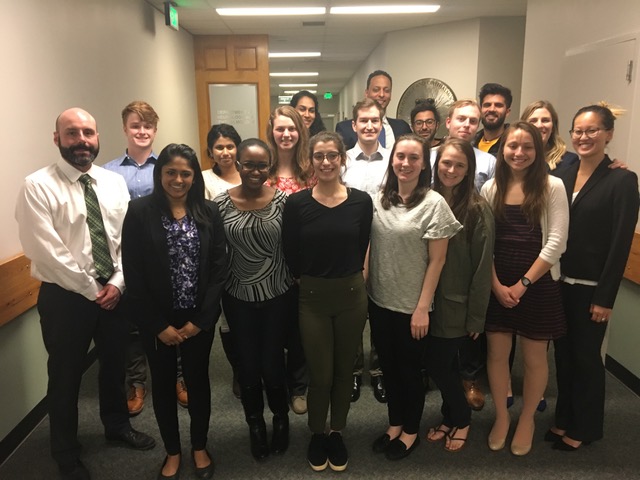
MSRJ is proud to announce its 2019-2020 Executive Board members!  Congratulations to Kathleen Louis-Gray, Eve Pourzan, Michael Dryden, Ninette Musili, Maria Rich, Antara Afrin, Christopher Dextras, and Jacob Purcell for being elected to their respective positions.  They are working hard to publish evidence-based and peer-reviewed research submitted by medical students across the globe.  We are excited to see how MSRJ will grow under their supervision!

Meet our staff:

Kathleen Louis-GrayExecutive Editor-In-Chief – is a 4th year medical student at MSU College of Human Medicine at the Traverse City Campus. She earned her Ph.D. in Neuroscience from Michigan State University, and studied thalamic synaptic plasticity in a rat model using electrophysiological techniques. She is currently in the Rural Community Health Program in Traverse City. There, she is involved in research regarding diet interventions and polysubstance use during pregnancy in a rural population. She has been an active member of MSRJ since 2016, helped to organize the MSRJ Elective, served as the M3 Executive Editor in the past. She is interested in neurology, and would like to strongly integrate clinical research with her future specialty.

Eve Pourzan M4 Executive Editor –  is a 3rd year medical student and is a prior 2017-2018 MSRJ Elective Organizer, and 2018-2019 Junior Editor Coordinator. She received her Bachelor’s in Politics from UCSC, and worked for the Michigan Legislature before pursuing medicine. Eve has lab experience investigating cell signaling in estrogen-positive breast cancer and systemic sclerosis. She is interested in pursuing anesthesiology.

Michael DrydenM3 Executive Editor – is a 3rd year medical student at MSU College of Human Medicine heading to the Traverse City campus in the coming months. He grew up in Petoskey, Michigan before heading to the University of Michigan where he completed his B.S. in Neuroscience. He then attended Eastern Michigan University where he received his M.S. in Cell and Molecular Biology with a focus in Behavioral Neuroscience. While there he studied mouse olfactory behavior and olfactory bulb plasticity while developing new equipment and methodology. He is currently interested in both Emergency Medicine and Neurology, and when not studying or on the wards, you will most likely find him outdoors either riding his bikes or spending time on the water.

Ninette MusiliSenior Editor Coordinator – Ninette is a rising 3rd year medical student at Michigan State University College of Human Medicine. She grew up in Ann Arbor, Michigan and earned her B.S. in Biomolecular Science from the University of Michigan. She is currently interested in maternal health and child disparities.

Maria RichJunior Editor Coordinator – Maria is a 2nd year medical student at Michigan State University College of Human Medicine. She grew up in Grand Rapids, Michigan and received her B.A. in Biology from Kalamazoo College where she enjoyed studying abroad in Quito, Ecuador and playing varsity soccer. Prior to starting medical school, she worked as a Clinical Research Coordinator with the BeatCC Pediatric Oncology Research Team. At this point in her medical education, she is excited about pediatrics, genetics, and palliative care.

Antara AfrinSecretary – Antara is a second year medical student at the Michigan State University College of Human Medicine, Grand Rapids campus. She grew up in Detroit, Michigan and received her B.S. in Biomolecular Science from the University of Michigan in Ann Arbor. During the summer prior to medical school, Antara worked with the research team of Dr. Mona Hanna-Attisha at the Pediatric Public Health Initiative to understand the effects of lead on development of children who were in utero at the height of the Flint Water Crisis. Antara’s current research interests are pediatrics, surgery, and neurodevelopmental disorders.

Christopher DextrasTreasurer – Chris is a third year medical student at Michigan State University’s College of Human Medicine. He grew up in Frederick, Maryland and received a B.S. in cell biology and molecular genetics from the University of Maryland. Prior to medical school, he worked at the NIH doing preclinical drug discovery research. He is currently interested in internal medicine.

Jacob PurcellPublic Relations – Jacob is an M2 at the Grand Rapids campus. Originally from Tampa, Florida, Jacob played collegiate baseball and earned his B.S. from Montreat College with a major in Biology and minor in Chemistry. He moved back to Tampa for a gap year and earned a graduate certificate in Clinical Investigation from the University of South Florida. He worked as a clinical research assistant at Moffitt Cancer Center and Morton Plant Hospital in the department of radiation oncology researching breast cancer genetics and radiation therapy outcomes. He also serves on the executive board for the MSUCHM-GR Surgery Interest Group and oversees undergraduate outreach events. His personal research interests include surgery, sports medicine, physical medicine & rehabilitation, and genetics.


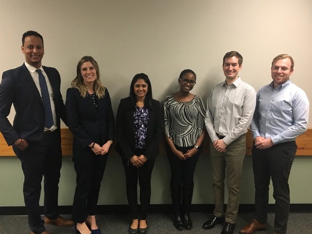
E-Board members (left to right) Eve Pourzan, Kathleen Louis-Gray, Ninette Musili, Christopher Dextras, and Michael Dryden.

Volume 7: Spring 2019 Issue

MSRJ Vol. 7 Spring 2019

The Spring 2019 issue is hot off the press! Click on the cover art to view and share the full issue!

Thank you to our artist, authors, student and faculty reviewers, and our E-Board for all your hard work and dedication to advancing evidence-based medicine!

Interested in a print copy? email us at @: contact@msrj.org

Malignant Chondroid Syringoma of the Foot – A Case Report

Authors: Megan Masten, MS41*, Raouf Mikhail, MD2

Author Affiliations:

1Michigan State University College of Human Medicine, Flint, Michigan, United States
2Surgical Oncologist, Hurley Medical Center, Flint, Michigan

Full Text Article PDF

*Corresponding Author: Megan Masten; mastenme@msu.edu

Key Words: Malignant; chondroid; syringoma; foot; adnexal cancer; cutaneous tumor

Abstract:

Background: This case report is about a very rare tumor – a malignant chondroid syringoma. The objective of this piece is to review both the case presented along with the current literature on cutaneous adnexal tumors.

Case Presentation: The patient is a 73-year-old Caucasian female with a past medical history of treated colon and breast cancer who presented with a 2-year history of a slow-growing, painful cutaneous lesion on the medial aspect of her right foot. The patient presented to her primary care physician (PCP) for right foot pain, which was attributed to bunions. The PCP encouraged the patient to see a podiatrist for this issue. Upon presentation to the podiatrist, the patient had a right foot biopsy. The pathology report showed a mixed malignant chondroid syringoma with positive margins. A re-excision to ensure complete removal was recommended. The patient presented to surgical oncology and subsequently she underwent complete excision of the right foot mass. At the time of her last visit, 7 months postoperatively, the patient continued to have issues with wound healing and continuous drainage of her surgical wound.

Discussion: This case differs from much of the current literature surrounding cutaneous adnexal tumors as it is a malignant chondroid syringoma of the foot, which is exceedingly rare. There are only three other published case reports of similar malignancies in similar places. This case study is important due to the uniqueness of the case. This case serves as a reminder of the importance of biopsy for diagnosis prior to management, as it is unlikely that such rare soft tissue tumors can be diagnosed without biopsy.

Conclusion: The take away lesson of the case is that it is important to biopsy unknown masses, and to have follow up with specific specialists.

Published: Spring, 2019

References:

1. Martinez SR, Barr KL, Canter RJ. Rare tumors through
the looking glass: an examination of malignant cutaneous adnexal tumors. Arch Dermatol 2011; 147(9): 1058–62.
doi: 10.1001/archdermatol.2011.229

2. Malik R, Saxena A, Kamath N. A rare case of malignant chondroid syringoma of scalp. Indian Dermatol Online
J 2013; 4(3): 236–8.

3. Mayur K, Neha M, Rajiv K, Shubhada K. Malignant chondroid syringoma of thigh with late metastasis to lung:
a very rare case report. Indian J Pathol Microbiol 2017;
60(3): 428–30.

4. Shashikala P, Chandrashekhar HR, Sharma S, Suresh KK. Malignant chondroid syringoma. Indian J Dermatol Venereol Leprol 2004; 70: 175–6.

5. American Joint Committee on Cancer TNM staging system for cutaneous squamous cell carcinoma. AJCC cancer staging manual. 7th ed. New York: Springer; 2010.

6. Lu H, Chen L, Chen Q, Shen H, Liu Z. A rare large cutaneous chondroid syringoma involving a toe: a case report. Medicine 2018; 97(5): e9825.

7. Madi K, Attanasio A, Cecunjanin F, Garcia R, Vidershayn A, Lucido, J. Chondroid syringoma of the foot: a rare diagnosis.
J Foot Ankle Surg 2016; 55: 373–8.

8. Sundling R, Logan D. Chondroid syringoma: a case report in the foot and ankle. Foot Ankle Specialist 2016; 10: 167–9.

9. Kazakov DV, McKee PH, Michal M, Kacerovska D. Cutaneous adnexal tumors. 1st ed. Philadelphia, PA: Lippincott Williams & Wilkins Health; 2012.

10. Conill C, Toscas I, Morilla I, Mascaró JM. Radiation therapy as a curative treatment in extraocular sebaceous carcinoma. Br J Dermatol 2003; 149(2): 441–2.

11. Duke WH, Sherrod TT, Lupton GP. Aggressive digital papillary adenocarcinoma (aggressive digital papillary adenoma and adenocarcinoma revisited). Am J Surg Pathol 2000; 24(6): 775–84. 12. Tolkachjov SN, Hocker TL, Camilleri MJ, Baum CL. Mohs micrographic surgery in the treatment of trichilemmal

carcinoma: the Mayo Clinic experience. J Am Acad Dermatol 2015; 72(1): 195.

13. Hamman M, Jiang S. Management of Trichilemmal Carcinoma: an update and comprehensive review of the literature. Dermatol Surg 2014; 40(7): 711–17.

14. Metzler G, Schaumburg-Lever G, Hornstein O, Rassner G. Malignant chondroid syringoma: immunohistopathology. Am J Dermatopathol 1996; 18: 83–9.

15. Webb JN, Stott WG. Malignant chondroid syringoma of the thigh. Report of a case with electron microscopy of the tumour. J Pathol 1975; 116: 43–6.

16. Mathiasen RA, Rasgon BM, Rumore G. Malignant chondroid syringoma of the face: a first reported case. Otolaryngol Head Neck Surg 2005; 133: 305–7.

17. Hirsch, P, Helwig, EB. Chondroid Syringoma. Arch. Derm. 1961; 84: 835–847.

Primary Intestinal Lymphangiectasia: A Case Report

Authors: Ridwaan Albeiruti1*, MD, Patrick Gleeson2, MD, Theodore Kelbel3, MD, Tracy Fausnight, MD3

Author Affiliations:

1Department of Medicine, West Virginia University, Morgantown, WV; Department of Internal Medicine, College of Human Medicine, Michigan State University, Grand Rapids, MI, USA; 2Department of Internal Medicine, Temple University, Philadelphia, PA, USA; 3Helen DeVos Children’s Hospital, Spectrum Health, Grand Rapids, MI, USA

Full Text Article PDF

*Corresponding Author: Ridwaan Albeiruti; albeiru4@msu.edu

Key Words: primary intestinal lymphangiectasia; Waldmann’s disease; protein-losing enteropathy

Abstract:

Primary intestinal lymphangiectasia (Waldmann’s disease) is a rare protein-losing enteropathy which is mostly seen in young children. A 22-month-old male baby presented with a 1-week history of abdominal distension, chronic loose stools, recurrent ear infections, and failure to thrive. He had edematous eyelids and non-pitting edema of his hands and feet. The patient was diagnosed via endoscopic visualization and biopsy of the lymphangiectasia in the small bowel. He was managed through dietary restriction with a high-protein, low-fat diet. The patient subsequently had resolution of the diarrhea and an increase in albumin and total protein on labs. We describe a rare case of primary intestinal lymphangiectasia and highlight its clinical presentation, diagnosis, and treatment.

Published: Spring, 2019

References:

1. Vignes S, Bellanger J. Primary intestinal lymphangiectasia (Waldmann’s disease). Orphanet J Rare Dis 2008; 3: 5.
doi: 10.1186/1750-1172-3-5

2. Wen J, Tang Q, Wu J, Wang Y, Cai W. Primary intestinal lymphangiectasia: four case reports and a review of the literature. Dig Dis Sci 2010; 55(12): 3466–72. doi: 10.1007/ s10620-010-1161-1

3. Hokari R, Kitagawa N, Watanabe C, Komoto S, Kurihara C, Okada Y, et al. Changes in regulatory molecules for lymphangiogenesis in intestinal lymphangiectasia with enteric protein loss. J Gastroenterol Hepatol 2008; 23(7 Pt 2): e88–95. doi: 10.1111/j.1440-1746.2007.05225.x

4. Katoch P, Bhardwaj S. Lymphangiectasia of small intestine presenting as intussusception. Indian J Pathol Microbiol 2008; 51(3): 411–12.

5. Dierselhuis MP, Boelens JJ, Versteegh FG, Weemaes C, Wulffraat NM. Recurrent and opportunistic infections in children with primary intestinal lymphangiectasia. J Pediatr Gastroenterol Nutr 2007; 44(3): 382–5. doi: 10.1097/01. mpg.0000233192.77521.2f

6. Ingle SB, Hinge Ingle CR. Primary intestinal lymphangiectasia: minireview. World J Clin Cases 2014; 2(10): 528–33. doi: 10.12998/wjcc.v2.i10.528

7. Xinias I, Mavroudi A, Sapountzi E, Thomaidou A, Fotoulaki M, Kalambakas A, et al. Primary intestinal lymphangiectasia: is it always bad? Two cases with different outcome. Case Rep Gastroenterol 2013; 7(1): 153–63. doi: 10.1159/000348763

Comparing Student Satisfaction with Traditional and Modular Group Peer-Tutoring Session

Authors:

Jeff Cross, MD1, Rodney Nyland PhD2, Sarah Lerchenfeldt, PharmD, BCPS, BCOP3

Author Affiliations:

1Oakland University William Beaumont School of Medicine, Rochester, MI, USA
2Department of Organizational Leadership, School of Education and Human Services, Oakland University, Rochester, MI, USA
3Department of Foundational Medical Studies, Oakland University William Beaumont School of Medicine, Rochester, MI, USA

Full Text Article PDF

*Corresponding Author: Jeff Cross; Jcross818@gmail.com

Key Words: education; medical; teaching; tutoring

Abstract:

Background: Our allopathic medical school has utilized a peer-tutoring program since inception in 2011, where second-year medical students teach first-year students in 2-h lecture-style review sessions. In 2015, an alternative format was implemented using four, repeating 30-min modules. This study was designed to compare student satisfaction with both approaches.

Methods: An online survey was emailed to students graduating in 2018 (n = 97) and 2019 (n = 127).

Results: A total of 72 (32.6%) responding students were included in the study, 35 from the class of 2018 (Co2018) and 37 from the class of 2019 (Co2019). Fewer Co2018 students, who received traditional instruction, were ‘very satisfied with the session timing’ compared with Co2019 students, who received the modular format (proportion difference: 0.42; P < 0.001, 95% confidence interval [CI] [0.21–0.63]). Co2018 students were more likely than Co2019 students to stop attending because their time was better utilized another way (proportion difference: 0.22; P = 0.054, 95% CI [-0.003 to 0.45]).

Conclusions: Students preferred the session length and timing of the modular format. Future studies are warranted to evaluate the effectiveness of this approach.

Published: Spring, 2019

References:

1. Benè KL, Bergus G. When learners become teachers:
a review of peer teaching in medical student education.
Fam Med 2014; 46(10): 783–7.

2. Sobral DT. Cross-year peer tutoring experience in a medical school: conditions and outcomes for student tutors. Med Educ 2002; 36(11so): 1064–70. doi: 10.1046/j.1365-2923.2002. 01308.x

3. Buckley S, Zamora J. Effects of participation in a cross year peer tutoring programme in clinical examination skills on volunteer tutors’ skills and attitudes towards teachers and teaching. BMC Med Educ 2007; 7(1): 20. doi: 10.1186/ 1472-6920-7-20

4. Yu TC, Wilson NC, Singh PP, Lemanu DP, Hawken SJ, Hill AG. Medical students-as-teachers: a systematic review of peer- assisted teaching during medical school. Adv Med Educ
Pract 2011; 2: 157–72. doi: 10.2147/amep.s14383

5. Lockspeiser TM, O’Sullivan P, Teherani A, Muller J. Understanding the experience of being taught by peers:
the value of social and cognitive congruence. Adv Heal Sci Educ 2008; 13(3): 361–72. doi: 10.1007/s10459-006-9049-8

6. Ten Cate O, Durning S. Peer teaching in medical education: twelve reasons to move from theory to practice. Med Teach 2007; 29(6): 591–9. doi: 10.1080/01421590701606799

7. Santee J, Garavalia L. Peer tutoring programs in health professions schools. Am J Pharm Educ 2006; 70(3): 70.
doi: 10.5688/aj700370

8. Swindle N, Wimsatt L. Development of peer tutoring services to support osteopathic medical students’ academic success. J Am Osteopath Assoc 2015; 115(11): e14–19. doi: 10.7556/jaoa.2015.140

9. Brown G, Manogue M. AMEE medical education guide
no. 22: refreshing lecturing: a guide for lecturers. Med Teach 2001; 23(3): 231–44. doi: 10.1080/01421590120043000